. 2010 Feb 11;463(7282):813-7.
doi: 10.1038/nature08699.

Human host factors required for influenza virus replication

Affiliations

Human host factors required for influenza virus replication

Renate König et al. Nature. .

Abstract

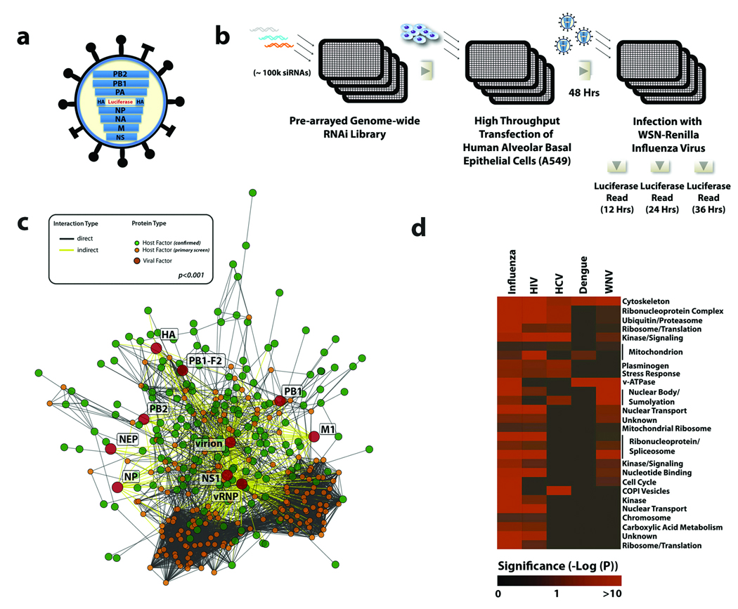
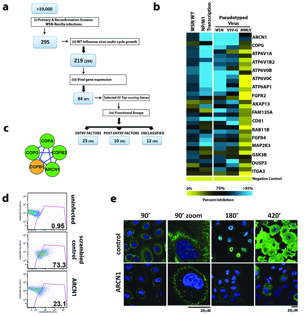
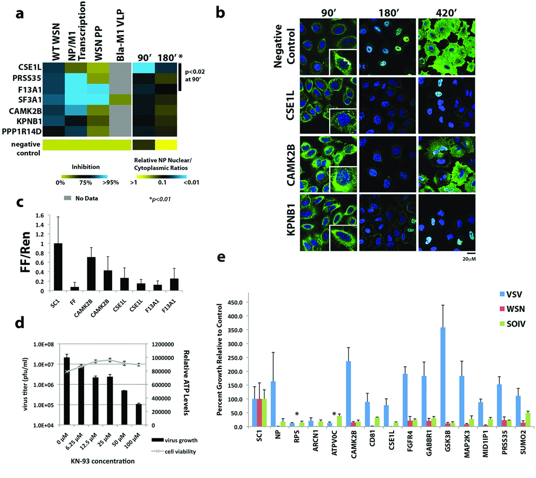
Influenza A virus is an RNA virus that encodes up to 11 proteins and this small coding capacity demands that the virus use the host cellular machinery for many aspects of its life cycle. Knowledge of these host cell requirements not only informs us of the molecular pathways exploited by the virus but also provides further targets that could be pursued for antiviral drug development. Here we use an integrative systems approach, based on genome-wide RNA interference screening, to identify 295 cellular cofactors required for early-stage influenza virus replication. Within this group, those involved in kinase-regulated signalling, ubiquitination and phosphatase activity are the most highly enriched, and 181 factors assemble into a highly significant host-pathogen interaction network. Moreover, 219 of the 295 factors were confirmed to be required for efficient wild-type influenza virus growth, and further analysis of a subset of genes showed 23 factors necessary for viral entry, including members of the vacuolar ATPase (vATPase) and COPI-protein families, fibroblast growth factor receptor (FGFR) proteins, and glycogen synthase kinase 3 (GSK3)-beta. Furthermore, 10 proteins were confirmed to be involved in post-entry steps of influenza virus replication. These include nuclear import components, proteases, and the calcium/calmodulin-dependent protein kinase (CaM kinase) IIbeta (CAMK2B). Notably, growth of swine-origin H1N1 influenza virus is also dependent on the identified host factors, and we show that small molecule inhibitors of several factors, including vATPase and CAMK2B, antagonize influenza virus replication.


Figures

Figure 1
Figure 1. A Genome-wide RNAi Screen for Influenza Virus Host Cellular Factors
(a) A schematic of the recombinant WSN-Ren virus showing the HA segment modified to express Renilla luciferase but maintaining the HA packaging sequences. (b) An arrayed genome-wide RNAi library (100,000 siRNAs targeting over 19,000 human genes) was transfected into A549 cells. Cells were subsequently infected with WSN-Ren and virus replication was monitored by measuring luciferase activities at the indicated times. (c) A highly significant (p<0.001 based on permutation test) host-pathogen interaction map for influenza virus containing 4,266 interactions between 181 confirmed influenza virus-host cellular factors (green circles), 10 influenza virus-encoded proteins or complexes (red circles), and an additional 184 cellular proteins (orange circles). (d) Predicted complexes from the human interactome used by influenza virus, HIV, HCV, dengue virus, and West Nile virus (based upon results reported here and previous screens–, –12, 28). Intensity of red colour indicates the significance of enrichment (based on hypergeometric p-values) for proteins required by a virus within a complex.
Figure 2
Figure 2. Identification of Host Factors Involved in Influenza Virus Entry
(a) Illustration of the screen progression from primary genome-wide analysis to the identification of factors involved in entry and post-entry steps in the virus life cycle. The number of confirmed genes and number of genes tested at each stage (in parentheses) are indicated. (b) The relative effects of gene depletion (2 siRNAs/gene) on infection of luciferase-encoding HIV particles pseudotyped with WSN, VSV or MMLV envelopes (right panel). Effects of RNAi upon wild-type WSN virus replication and transcription of viral NP and M1 genes are also shown (left panel). Inhibition in each assay is shown as a continuum of blue (high inhibition) to yellow (low inhibition). (c) The endosomal coat protein complex (COPI) was identified as one of seventeen biochemical modules (MCODE clusters) likely to be important for influenza virus entry (see Figure 1c for legend; also Supplementary Figure S5). (d) Infection of siRNA-transfected A549 cells with influenza virus VLPs carrying a beta-lactamase (Bla-M1) fusion protein. The percentages of cells containing detectable cytoplasmic beta-lactamase activity are indicated. (e) Cells depleted of ARCN1 and infected with wild-type WSN virus were fixed and stained for NP (green) and nuclei (blue) at the indicated times and analyzed by confocal microscopy. The enlarged images at 90min post-infection indicate the lack of incoming RNP complexes in the nucleus in cells depleted of ARCN1.
Figure 3
Figure 3. Characterization of Factors in Post-Entry Replication Events and Conserved Requirement by Different Influenza Viruses
(a) The impact of host factor depletion on the nuclear localization of viral NP protein at 90 and 180 minutes after A/WSN/33 virus infection is shown (right panel). Significant effects (p<0.01 based on Welch T-test) are seen at 180min with all genes and with CSE1L, PRSS35, F13A1 (p<0.02) at 90min. Levels of virus replication (WT WSN), viral gene (NP/M1) transcription and entry of WSN pseudotyped particles or Bla-M1 VLPs in cells lacking these factors are shown in the left panel. Values relative to negative controls (bottom row) are depicted in a continuum of blue (>95% inhibition) to yellow (little or no inhibition). (b) Confocal imaging of influenza virus NP protein localization at the indicated times following A/WSN/33 virus infection in cells depleted of CSE1L, CAMK2B and KPNB1. Arrows in the 90’ inset indicate nuclear RNPs. (c) The effects of host factor depletion on replication of an influenza virus mini-genome firefly reporter. The normalized fold reduction of firefly luciferase for each gene is shown relative to the scrambled siRNA control (SC1) +/− standard deviation. All reductions are significant (p<0.05) by Student’s T-test. (FF=firefly luciferase siRNA). (d) KN-93, a selective inhibitor of CAMK2B, inhibits A/WSN/33 influenza viral replication in a dose-dependent manner in MDCK cells, without affecting cell viability (ATP levels). Mean titers +/− standard deviation of triplicate samples are shown. (e) A549 cells were transfected with siRNAs targeting the indicated genes and subsequently infected with influenza A/WSN/33 virus, swine-origin influenza A/Netherlands/602/2009 (H1N1) virus (SOIV) or VSV. Virus growth is shown as the average percent relative to the scrambled siRNA control (SC1) +/− standard deviation. *below level of detection (1 × 10⁴ pfu/ml). NP=siRNA for influenza A virus NP, RPS=siRNA for RPS27A.

Similar articles

Cited by

References

    1. Palese P, Shaw ML. Orthomyxoviridae: The Viruses and Their Replication. In: Knipe DM, Howley PM, editors. Fields Virology. 5th Edition. Vol. 2. Philadelphia: Lippincott Williams & Wilkins; 2007. pp. 1647–1689.
    1. Wang TT, Palese P. Unraveling the mystery of swine influenza virus. Cell. 2009;137:983–985. - PubMed
    1. De Clercq E. Antiviral agents active against influenza A viruses. Nat Rev Drug Discov. 2006;5:1015–1025. - PMC - PubMed
    1. Layne SP, Monto AS, Taubenberger JK. Pandemic influenza: an inconvenient mutation. Science. 2009;323:1560–1561. - PMC - PubMed
    1. Brass AL, et al. Identification of host proteins required for HIV infection through a functional genomic screen. Science. 2008;319:921–926. - PubMed

Publication types

MeSH terms

Substances