

Review
doi: 10.1038/nrm2815.
Mechanisms guiding primordial germ cell migration: strategies from different organisms
Affiliations
- PMID: 20027186
- PMCID: PMC4521894
- DOI: 10.1038/nrm2815
Item in Clipboard
Review
Mechanisms guiding primordial germ cell migration: strategies from different organisms
Nat Rev Mol Cell Biol.
2010 Jan.
Abstract
The regulated migration of cells is essential for development and tissue homeostasis, and aberrant cell migration can lead to an impaired immune response and the progression of cancer. Primordial germ cells (PGCs), precursors to sperm and eggs, have to migrate across the embryo to reach somatic gonadal precursors, where they carry out their function. Studies of model organisms have revealed that, despite important differences, several features of PGC migration are conserved. PGCs require an intrinsic motility programme and external guidance cues to survive and successfully migrate. Proper guidance involves both attractive and repulsive cues and is mediated by protein and lipid signalling.
Figures
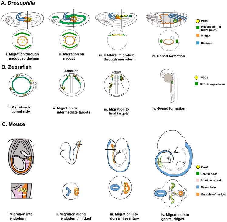
a | Drosophila melanogaster. i. After specification Primordial germ cells (PGCs) are carried into the embryo by the midgut primordium. PGCs polarize and migrate through the midgut epithelium at stages 9–10 (∼4.5h After Egg Laying (AEL)). ii. PGCs reorient on midgut towards the mesoderm at stage 10 (∼5h 10m AEL). iii. PGCs migrate bilaterally towards the somatic gonadal precursors (SGPs) at stage 11 (∼7h AEL). iv. PGCs associate with SGPs and coalesce to form the embryonic gonad. Lateral views (top) and transverse sections (bottom). b | Zebrafish. i. Following specification at four random locations, PGCs migrate dorsally (animal pole view; the animal pole refers to the portion of the blastula embryo that differentiates into mesoderm and ectoderm). ii. At gastrulation, 4.5 hours post-fertilization (hpf), PGCs follow expression of stromal derived factor 1a (SDF-1a). Somites 1-3 act as intermediate targets (lateral view, left side) at 10.5 hpf. iii. PGCs migrate towards the final target tissue at somites 8-10 (frontal view) at 13hpf. iv. At 24 hpf, PGCs coalesce with the somatic cells of the gonad (lateral view, left side). c | Mouse. i. PGCs, specified in proximal epiblast, migrate from the primitive streak to the endoderm (future hindgut) at embryonic day 7.5 (E7.5). Closeup shown on bottom. ii. At E8, PGCs migrate along the endoderm. iii, At E9.5, PGCs migrate bilaterally towards the dorsal body wall. iv. At E10.5, PGCs reach the genital ridges to form the embryonic gonad. Lateral views (top) and transverse sections (bottom). Adapted from Starz-Gaiano and Lehmann (2001) and Santos and Lehmann (2004), .
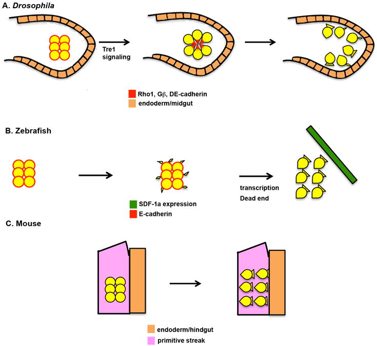
a | Drosophila melanogaster. I. At early stage 9 (∼4h After Egg Laying (AEL)), germ cells are tightly clustered in the midgut pocket. Primordial germ cells (PGCs) are not polarized at this stage and show little interaction with the midgut primordium. E-Cadherin, the small GTPase Rho1 and Gβ proteins are present uniformly at the cell periphery. Trapped in endoderm 1 (Tre1) signalling leads to the polarization of the PGCs, which take on a radial organization with the tails of the cells facing the inside of the cluster and the leading edges facing the midgut primordium. E-Cadherin, Rho1 and Gβ are redistributed to the tails of the cells. Next, the PGCs lose adhesion to each other and begin to extend cellular protrusions towards the epithelial cells of the midgut. b | Zebrafish. At specification, PGCs have a smooth, round morphology and do not posses migratory activity (3 hours post-fertilization (hpf)). PGCs begin to randomly extend small cellular protrusions in multiple directions at 3.5hpf. These protrusions disappear during mitosis. At 4.5hpf, PGCs become polarized, individualize and extend broad protrusions at the leading edge. This step is dependent on transcription and the Dead End protein, and is necessary for the cells to respond to stromal derived factor 1a (SDF-1a, also known as CXCL12a) chemokine signalling. c | Mouse. Following specification in the posterior primitive streak (embryonic day 7.5), PGCs have a smooth, round morphology. PGCs acquire a polarized morphology prior to initiating their migration into the endoderm. The molecular mechanisms regulating this polarization are not understood.

a | Drosophila melanogaster. i. The G-protein coupled receptor (GPCR) Trapped in endoderm 1 (Tre1) regulates transepithelial migration of primordial germ cells (PGCs) through the midgut. Tre1 might regulate Rho1, triggering cytoskeletal changes necessary for migration. ii. Wunen and Wunen2 (Wun and Wun2) regulate migration into the mesoderm. Wun and Wun2 are expressed at sites that PGCs avoid, such as the ventral midgut, and in PGCs. Data suggest that Wun and Wun2 hydrolyze an extracellular phospholipid that functions as a PGC attractant and survival factor. iii. PGCs are attracted to somatic gonad precursors (SGPs) by the 3-hydroxy-3-methylglutaryl coenzyme A reductase (Hmgcr) pathway, which adds a geranyl-geranyl (GG) group to a putative chemoattractant. Multidrug resistance 49 (Mdr49), an ABC transporter, is required for chemoattractant secretion. b | Zebrafish. PGCs expressing the GPCR Chemokine (CXC motif) Receptor 4b (CXCR4b) migrate towards the CXCR4b ligand, stromal derived factor 1a (SDF-1a), secreted by somatic cells. Another somatically-expressed GPCR, CXCR7b, promotes internalization and degradation of SDF-1a, which might lead to proper gradient formation and precise targeting of PGCs. Following PGC migration to an intermediate target, a new group of distant somatic cells begins expressing SDF-1a, directing PGCs to new targets. c | Mouse. PGC migration to the genital ridges is controlled by the GPCR CXCR4 and its ligand SDF-1. SDF-1 is expressed by the somatic cells of the genital ridge and PGCs express CXCR4. Integrin β1 is also required for this step. PGC motility and survival requires the receptor tyrosine kinase c-Kit and its ligand Steel. Steel is expressed by somatic cells surrounding PGCs throughout migration.
References
-
- Ridley AJ, et al. Cell migration: integrating signals from front to back. Science. 2003;302:1704–9. - PubMed
-
- Charras G, Paluch E. Blebs lead the way: how to migrate without lamellipodia. Nat Rev Mol Cell Biol. 2008;9:730–6. - PubMed
-
- Webb DJ, Zhang H, Horwitz AF. Cell migration: an overview. Methods Mol Biol. 2005;294:3–11. - PubMed
-
- Franz CM, Jones GE, Ridley AJ. Cell migration in development and disease. Dev Cell. 2002;2:153–8. - PubMed
Publication types
MeSH terms
Grants and funding
LinkOut - more resources
Full Text Sources
Other Literature Sources
