. 2009 Dec 21;4(12):e8268.
doi: 10.1371/journal.pone.0008268.

Vitamin B12-impaired metabolism produces apoptosis and Parkinson phenotype in rats expressing the transcobalamin-oleosin chimera in substantia nigra

Affiliations

Vitamin B12-impaired metabolism produces apoptosis and Parkinson phenotype in rats expressing the transcobalamin-oleosin chimera in substantia nigra

Carlos Enrique Orozco-Barrios et al. PLoS One. .

Abstract

Background: Vitamin B12 is indispensable for proper brain functioning and cytosolic synthesis of S-adenosylmethionine. Whether its deficiency produces effects on viability and apoptosis of neurons remains unknown. There is a particular interest in investigating these effects in Parkinson disease where Levodopa treatment is known to increase the consumption of S-adenosylmethionine. To cause deprivation of vitamin B12, we have recently developed a cell model that produces decreased synthesis of S-adenosylmethionine by anchoring transcobalamin (TCII) to the reticulum through its fusion with Oleosin (OLEO).

Methodology: Gene constructs including transcobalamin-oleosin (TCII-OLEO) and control constructs, green fluorescent protein-transcobalamin-oleosin (GFP-TCII-OLEO), oleosin-transcobalamin (OLEO-TCII), TCII and OLEO were used for expression in N1E-115 cells (mouse neuroblastoma) and in substantia nigra of adult rats, using a targeted transfection with a Neurotensin polyplex system. We studied the viability and the apoptosis in the transfected cells and targeted tissue. The turning behavior was evaluated in the rats transfected with the different plasmids.

Principal findings: The transfection of N1E-115 cells by the TCII-OLEO-expressing plasmid significantly affected cell viability and increased immunoreactivity of cleaved Caspase-3. No change in propidium iodide uptake (used as a necrosis marker) was observed. The transfected rats lost neurons immunoreactive to tyrosine hydroxylase. The expression of TCII-OLEO was observed in cells immunoreactive to tyrosine hydroxylase of the substantia nigra, with a superimposed expression of cleaved Caspase-3. These cellular and tissular effects were not observed with the control plasmids. Rats transfected with TCII-OLEO expressing plasmid presented with a significantly higher number of turns, compared with those transfected with the other plasmids.

Conclusions/significance: In conclusion, the TCII-OLEO transfection was responsible for apoptosis in N1E-115 cells and rat substantia nigra and for Parkinson-like phenotype. This suggests evaluating whether vitamin B12 deficit could aggravate the PD in patients under Levodopa therapy by impairing S-adenosylmethionine synthesis in substantia nigra.


Conflict of interest statement

Competing Interests: The authors have declared that no competing interests exist.

Figures

Figure 1
Figure 1. Expression of transcobalamin/oleosin chimeric proteins in transfected N1E-115 cells.
A: Transgenic expression in N1E-115 cells at 48 h after transfection using lipofectamine. The cells were transfected with one of the following plasmids: pCMV-TCII-OLEO coding for transcobalamin-oleosin (lane 1), pCMV-OLEO-TCII coding for oleosin-transcobalamin (lane 2), pCMV-TCII coding for transcobalamin II (lane 3), pCMV-OLEO coding for oleosin (lane 4), pCDNA3 (lane 5), and pCMV-GFP-TCII-OLEO coding for GFP-TCII-OLEO (lane 6). The housekeeping gene was β-actin. The size in bp of the amplified products was 1347 for transcobalamin-oleosin (TCII-OLEO), 1240 for oleosin-transcobalamin (OLEO-TCII), 551 for transcobalamin II (TCII), 275 for oleosin (OLEO), and 349 for β-actin. B: Western blotting of homogenate of N1E-115 cells transfected with the various plasmids. From lane 1 to 6: homogenates from cells transfected with pCMV-TCII-OLEO, pCMV-OLEO-TCII, pCMV-TCII, pCMV-OLEO, empty plasmid, pCMV-GFP-TCII-OLEO, respectively. C: Vitamin B12 binding capacity in transfected cells. 57Co-labeled Cobalamin (Cbl, ∼300 µCi per µg) was incorporated into culture medium (30,000 dpm/mL) for three days. The total amount of radioactivity taken by each cell lines was measured in pellets and supernatants. Mean and S.E.M. are indicated. D: Indirect immunofluorescence of TCII in N1E-115 cells transiently transfected with lipofectamine. The four constructs and the empty plasmid were tranfected in N1E-115 cells (1–5). The immunofluorescence was done with a goat polyclonal antibody to TCII and a donkey antigoat IgG fluorescein labeled. Cell nuclei were counterstained with Hoechst 33258. Calibration bars = 10 µm. E: Confocal analysis showing co-localization of the protein GFP-TCII-OLEO with endoplasmic reticulum in transfected N1E-115 cells. The cells were transfected with the plasmid pCMV-GFP-TCII-OLEO coding for GFP-transcobalamin-oleosin (GFP-TCII-OLEO), using lipofectamine. Cell nuclei were counterstained with Hoechst 33258 (1, 5). Co-localization was evidenced with fluorescence from GFP (2, 6), immuno-fluorescence with a mouse monoclonal antibody to the human golgin-97 (3) or a rabbit polyclonal antibody to calreticulin (7) and merge fluorescence (4,8). The secondary antibodies include a donkey IgG anti-mouse TRITC labeled or a donkey IgG anti-rabbit TRITC labeled. Calibration bars = 20 µm.
Figure 2
Figure 2. Viability and apoptosis of N1E-115 cells stably transfected with different plasmids.
The plasmids were pCMV-TCII-OLEO coding for transcobalamin-oleosin (TCII-OLEO), pCMV-OLEO-TCII coding for oleosin-transcobalamin (OLEO-TCII), pCMV-TCII coding for transcobalamin II (TCII), pCMV-OLEO coding for oleosin (OLEO), and pCDNA3. A: Cell viability and apoptosis among growth delay in proliferated state was monitored by the absorbance of formazan dye resulting from enzymatic metabolism of MTT by the mitochondrial dehydrogenase. The no reagent blank was MTT alone (0.5 mg/mL in culture medium). Western blot analysis of the temporal course of p53 in cells transfected with pCMV-TCII-OLEO was made by using a mouse anti-p53 antibody and a mouse anti-actin antibody, which were revealed with a goat anti-mouse IgG coupled to peroxidase. Cleaved Caspase-3 was identified by a rabbit anti-caspase-3 polyclonal antibody and a donkey anti-rabbit secondary antibody (labeled with peroxidase). B: Western blotting of cleaved Caspase-3 at Day 7 of proliferate state. The mean±S.E.M. were obtained from three independent experiments made in triplicate. Two-way ANOVA and Bonferoni post-test analyzed statistical differences from the control groups. *, P<0.05; **, P<0.01; ***, P<0.001.
Figure 3
Figure 3. Analysis of apoptosis in N1E-115 cells stably transfected with different plasmids.
The plasmids were pCMV-TCII-OLEO coding for transcobalamin-oleosin (TCII-OLEO), pCMV-OLEO-TCII coding for oleosin-transcobalamin (OLEO-TCII), pCMV-TCII coding for transcobalamin II (TCII), pCMV-OLEO coding for oleosin (OLEO), and pCDNA3. The immunofluorescence was done with a rabbit polyclonal antibody to cleaved Caspase-3 and a donkey antirabbit IgG fluorescein labeled. Before fixation, cells were incubated with 4 µM propidium iodide for 10 min. Cell nuclei were counterstained with Hoechst 33258. Calibration bars = 100 µm.
Figure 4
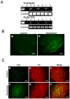
Figure 4. Expression of transcobalamin II/oleosin (TCII/OLEO) chimeric proteins in rats 60 days after transfection with the NTS-polyplex.
A: RT-PCR from the plasmid transcripts in the substantia nigra of rats. A group of rats (n = 3) was transfected with the plasmid pCMV-TCII-OLEO and another (n = 3) with the plasmid pCMV-OLEO-TCII. RT-PCR amplified a fragment of 380 bp for TCII-OLEO, a fragment of 394 for OLEO-TCII, and a fragment of 349 for β-actin, the internal control. Lane 1 corresponds to the amplified fragment from the plasmid (positive control). Lane 2 is a PCR in the absence of plasmid or cDNA (negative control). The amplified product from the transfected substantia nigra of each rat corresponds to the lanes 3, 5, and 7, and the lanes 4, 6, and 8 show the RT-PCR outcome from the non-transfected side. B: GFP immunofluorescence in the rat substantia nigra transfected with pCMV-GFP-TCII-OLEO. The pCMV-GFP-TCII-OLEO encodes for the fusion protein green fluorescent protein-transcobalamin-oleosin (GFP-TCII-OLEO). The immunofluorescence was done with a mouse monoclonal antibody to GFP and a donkey antimouse IgG fluorescein labeled. Representative micrographs of coronal section of control substantia nigra (1) and transfected substantia nigra (2) of the same rat are presented. Calibration bars = 100 µm. C: Double immunofluorescence against TCII and tyrosine hydroxylase (TH) in the substantia nigra of rats. The neurons were transfected with NTS-polyplex with pCMV-TCII-OLEO coding for transcobalamin-oleosin (TCII-OLEO). Slices from mesencephalon (40 µm) were immunostained at 7-day after transfection. The primary antibodies were a goat polyclonal anti-TCII and a mouse monoclonal anti-TH. The secondary antibodies were a donkey antigoat IgG fluorescein labeled and a donkey antimouse IgG rhodamine labeled. Representative micrographs of coronal section of control substantia nigra (1–3) and transfected substantia nigra (4–6) of the same rat are presented. Calibration bars = 50 µm.
Figure 5
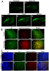
Figure 5. Apoptosis of tyrosine hydroxylase (TH) immunoreactive cells in the substantia nigra of rats transfected with several plasmids.
A: TH-immunoreactive neurons after transfection. The neurons were transfected with NTS-polyplex with one of the following plasmids, pCMV-TCII-OLEO coding for transcobalamin-oleosin (TCII-OLEO, 1), pCMV-OLEO-TCII coding for oleosin-transcobalamin (OLEO-TCII, 2), pCMV-TCII coding for transcobalamin II (TCII, 3), pCMV-OLEO coding for oleosin (OLEO, 4), and the pCDNA3, the empty plasmid (5). Mesencephalon slices (40 µm) were immunostained at 2-month after transfection with a mouse monoclonal antibody to TH and a donkey antimouse IgG fluorescein labeled. Representative micrographs of sagital section of the rat mesencephalon are presented. Calibration bars = 200 µm. B: Apoptosis in TH-immunoreactive neurons after transfection with the plasmid pCMV-TCII-OLEO. Representative micrographs of the substantia nigra (with double immunostaining at 15-day after transfection) are presented. The primary antibodies were a mouse monoclonal antibody to TH, and a rabbit polyclonal antibody to cleaved Caspase-3. The secondary antibodies included a donkey anti-mouse IgG FITC labeled (1 and 4), and a donkey anti-rabbit IgG rhodamine labeled (2 and 5). Representative micrographs of coronal section of control substantia nigra (1–3) and transfected substantia nigra (4–6) of the same rat are presented. Scale bars = 50 µm. C: Apoptosis in TH immunoreactive neurons expressing the TCII-OLEO chimera. Representative micrographs of the substantia nigra with triple immunostaining at 15-day after transfection. The primary antibodies were a mouse monoclonal antibody to TH, a goat polyclonal antibody to TCII, and a rabbit polyclonal antibody to cleaved Caspase-3. The secondary antibodies were a donkey anti-mouse IgG AMCA labeled (1 and 5), a donkey antigoat IgG fluorescein labeled (2 and 6), a donkey anti-rabbit IgG rhodamine labeled (3 and 7). Representative micrographs of coronal section of control substantia nigra (1–4) and transfected substantia nigra (5–8) of the same rat are presented. Scale bars = 50 µm.
Figure 6
Figure 6. Methamphetamine-induced turning behavior in rats transfected with several plasmids.
The plasmids were pCMV-TCII-OLEO coding for transcobalamin-oleosin (TO), pCMV-OLEO-TCII coding for oleosin-transcobalamin (OT), pCMV-TCII coding for transcobalamin II (T), pCMV-OLEO coding for oleosin (O), and pCDNA3 (P). The values are the mean±SEM of 3 animals per group. ** = Significantly different from control groups. P<0.01, repeated-measures two-way ANOVA and Bonferroni post-test.

References

    1. Russell JSR, Batten FE, Collier J. Subacute combined degeneration of the spinal cord. Brain. 1900;23:39–110.
    1. Scalabrino G. The multi-faceted basis of vitamin B12 (cobalamin) neurotrophism in adult central nervous system: Lessons learned from its deficiency. Prog Neurobiol. 2009;88:203–20. - PubMed
    1. Forges T, Monnier-Barbarino P, Alberto JM, Guéant-Rodriguez RM, Daval JL, et al. Impact of folate and homocysteine metabolism on human reproductive health. Hum Reprod Update. 2007;13:225–38. - PubMed
    1. Cheng H, Gomes-Trolin C, Aquilonius SM, Steinberg A, Löfberg C, et al. Levels of L-methionine S-adenosyltransferase activity in erythrocytes and concentrations of S-adenosylmethionine and S-adenosylhomocysteine in whole blood of patients with Parkinson's disease. Exp Neurol. 1997;145:580–5. - PubMed
    1. Fuso A, Seminara L, Cavallaro RA, D'Anselmi F, Scarpa S. S-adenosylmethionine/homocysteine cycle alterations modify DNA methylation status with consequent deregulation of PS1 and BACE and beta-amyloid production. Mol Cell Neurosci. 2005;28:195–204. - PubMed

Publication types

MeSH terms