. 2009 Dec 21;4(12):e8366.
doi: 10.1371/journal.pone.0008366.

Opposite effects of low and high doses of Abeta42 on electrical network and neuronal excitability in the rat prefrontal cortex

Affiliations

Opposite effects of low and high doses of Abeta42 on electrical network and neuronal excitability in the rat prefrontal cortex

Yun Wang et al. PLoS One. .

Abstract

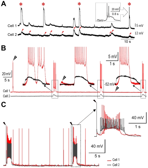
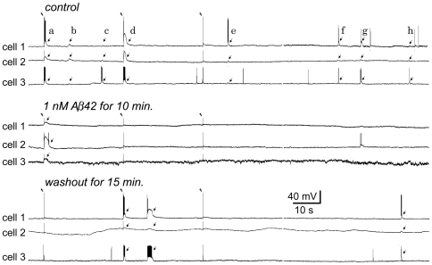
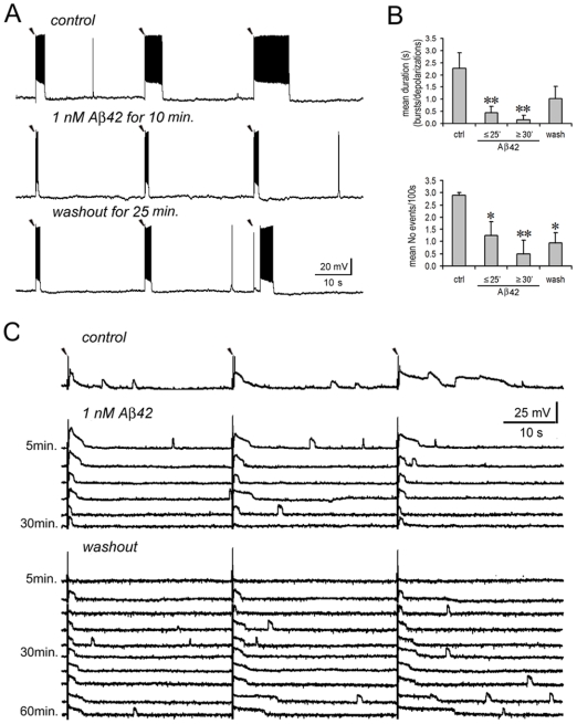
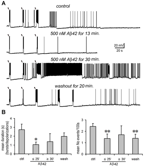
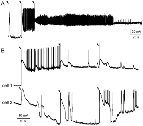
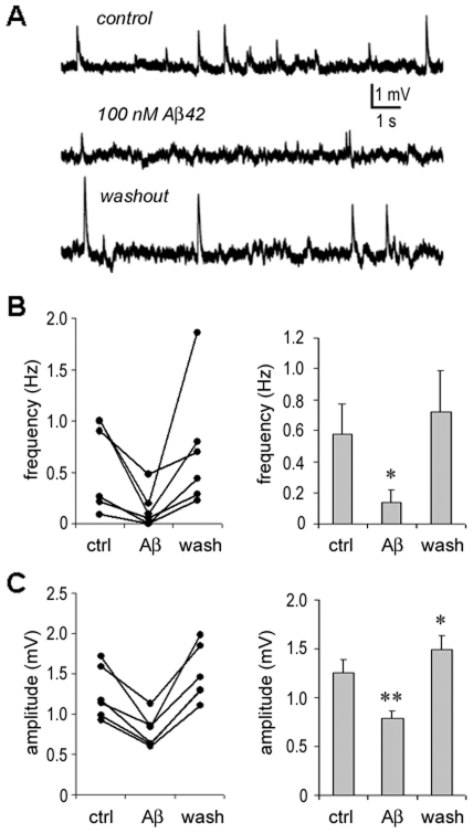
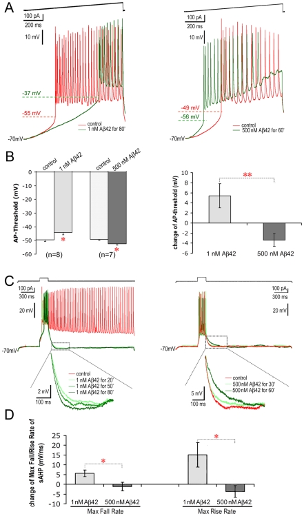
Changes in neuronal synchronization have been found in patients and animal models of Alzheimer's disease (AD). Synchronized behaviors within neuronal networks are important to such complex cognitive processes as working memory. The mechanisms behind these changes are not understood but may involve the action of soluble beta-amyloid (Abeta) on electrical networks. In order to determine if Abeta can induce changes in neuronal synchronization, the activities of pyramidal neurons were recorded in rat prefrontal cortical (PFC) slices under calcium-free conditions using multi-neuron patch clamp technique. Electrical network activities and synchronization among neurons were significantly inhibited by low dose Abeta42 (1 nM) and initially by high dose Abeta42 (500 nM). However, prolonged application of high dose Abeta42 resulted in network activation and tonic firing. Underlying these observations, we discovered that prolonged application of low and high doses of Abeta42 induced opposite changes in action potential (AP)-threshold and after-hyperpolarization (AHP) of neurons. Accordingly, low dose Abeta42 significantly increased the AP-threshold and deepened the AHP, making neurons less excitable. In contrast, high dose Abeta42 significantly reduced the AP-threshold and shallowed the AHP, making neurons more excitable. These results support a model that low dose Abeta42 released into the interstitium has a physiologic feedback role to dampen electrical network activity by reducing neuronal excitability. Higher concentrations of Abeta42 over time promote supra-synchronization between individual neurons by increasing their excitability. The latter may disrupt frontal-based cognitive processing and in some cases lead to epileptiform discharges.


Conflict of interest statement

Competing Interests: The authors have declared that no competing interests exist.

Figures

Figure 1
Figure 1. Recordings of synchronized responses between individual neurons in an electrical network of rat PFC.
A. Synchronized spontaneous depolarizations (marked with red arrow heads). Responses were recorded between two PCs (cell 1 and cell 2) under calcium-free conditions. The depolarizations of cell 1 lead to a few action potentials (APs) (for clarity, APs were truncated as marked with stars). B. Synchronized spontaneous bursts and depolarizations. Recording procedure was the same as in A. Bursts of cell 1 were synchronized with depolarizations of cell 2. The synchronized responses varied from having equal time courses up to 750 ms differences in duration (as shown in the insets, APs were truncated). Solitary or un-synchronized responses of cell 1 are indicated with double black arrow heads. C. Experimentally evoked synchronizing responses. Two neurons (cell 1 and cell2) were simultaneously recorded while two separated extracellular stimuli (20 µA for a duration of 2 ms; marked with single black arrow heads) were delivered locally at 30 s intervals. Bursting responses were provoked in both cells immediately after either of the stimulating artifacts. These bursts were synchronized yet had un-equal durations (see the inset). Following the evoked bursts, spontaneous APs continued to be captured from cell 1, but none from cell 2.
Figure 2
Figure 2. Low dose Aβ42 inhibits synchronized responses in a triple-neuron recording.
Under calcium-free conditions, three PCs were simultaneously recorded (cells 1–3). Three extracellular stimuli (30 µA, 2 ms duration) were delivered locally at 30 s intervals (marked with arrow heads). Thereafter the recording was continued for more than 100 s to capture spontaneous responses. This recording procedure was repeated for the conditions of pre-application, application and washout of Aβ. Upper panel: In the control condition, 2 (the 1st and 2nd) evoked responses (arrows a and d) and 6 spontaneous responses (arrows b,c, e–h) were synchronized between the triple-neuron assembly. The synchronized responses consist of bursts and depolarizations in cell 1 & cell 3 and only depolarizations in cell 2. Note that not all responses were synchronized. Middle panel: After 1 nM Aβ42 was applied for 10 min, both evoked and spontaneous responses were notably inhibited. Only 1 subthreshold response was evoked and synchronized among the three cells. Further, burst firing was lost in cell 1 & cell 3. Lower panel: After washout of Aβ42 for 15 min., the responses (1 evoked and 2 spontaneous) and their synchronicity tended to recover. Burst firing was also resumed in cells 1 and cell 3.
Figure 3
Figure 3. Low dose Aβ42 inhibits electrical network activities.
A. A representative experiment. Under calcium-free conditions, a PC was recorded while three extracellular stimuli (35 µA, 2 ms duration) were delivered in close proximity. The same recording procedure was repeated in pre-application, application and washout of Aβ. Following each stimulating artifact (marked with an arrow head), burst firing was provoked (upper panel). The bursts became briefer after 1 nM Aβ42 was applied (middle panel), and tended to recover on washout for 25 min. (lower panel). Note that the 3rd evoked burst in washout procedure occurred with a few seconds delay after the stimulating artifact. B. Statistical analysis. Upper panel: Compared with the control, the average duration of evoked responses was significantly reduced after 1 nM Aβ42 was applied for ≤25 min. (p = 0.010, n = 16), and appeared to be further reduced after prolonged applications for ≥30 min. (p = 0.009). On washout, the average duration recovered (Difference became insignificant compared with control, p = 0.07). Lower panel: Compared with the control, the average occurrence of evoked responses was significantly reduced after 1 nM Aβ42 was applied for ≤25 min. (p = 0.014), and even further reduced after the application for ≥30 min. (p = 0.009). On washout, this occurrence tended to recover but was still significantly lower than the control (p = 0.011). C. Another representative experiment. The experimental procedure is the same as in A (The extracellular stimulus was 30 µA for 2 ms; marked with arrow heads). In the control conditions, evoked and spontaneous depolarizations were visible following each stimulating artifact. After applying 1 nM Aβ42, the same recording was repeated for 30 min. in 5 min. intervals. Over time, evoked depolarizations gradually became narrower, and finally both evoked and spontaneous depolarizations vanished. Following washout, evoked depolarizations re-occurred and became gradually broader. Spontaneous depolarizations also reappeared and became more and more frequent. Note: “*”, p<0.05; “**”, p<0.01.
Figure 4
Figure 4. High dose Aβ42 has a biphasic effect on electrical network activities.
A. A representative experiment. Under calcium-free conditions, a PC was recorded after delivery of three extracellular stimuli. The bursts became briefer after 500 nM Aβ42 was applied for 13 min. (2nd panel). After Aβ was applied for ≥30 min., the responses became paradoxically stronger as evidenced by the broader bursts and tonic firing that followed the 3rd evoked burst (3rd panel). Spontaneous responses were also enhanced (followed the 2nd evoked burst in the 3rd panel). The excessive activity recovered on washout for 20 min. (lower panel). B. Statistical analysis: Compared with the control, the average duration (left panel) and occurrence (right panel) of evoked responses was significantly reduced after 500 nM Aβ42 was applied for ≤25 min. (duration: p = 0.017; occurrence: p = 0.002, n = 10, not including the cells that later showed tonic responses during prolonged application). The inhibition disappeared with Aβ applications for more than 30 min. (duration: p = 0.509; occurrence: p = 0.137). On washout, responses were similar to the control (duration: p = 0.332) but appeared less frequently (occurrence: p = 0.006). Note: “*”, p<0.05; “**”, p<0.01.
Figure 5
Figure 5. Tonic firing and supra-synchronization.
A. A tonic firing recorded from a PC. After 500 nM Aβ42 was applied for more than 30 min., tonic firing was induced in a PC immediately after the 2nd stimulus was delivered. The membrane potential thereafter was found to be depolarized to −20 mV for more than 10 min. (200 s recording was shown). B. Synchronization during and after tonic firing. In a PC-pair recording exposed to 500 nM Aβ42 for 30 min., a stimulating pulse in cell 1 initially provoked tonic AP firing. Its membrane potential became depolarized similar to A. This highly depolarized state remained stable without AP firing for more than 10 min. (not show all). Cell 2 initially experienced hyperpolarization. Nevertheless, almost all depolarizing responses, lasting for up to 10 s, showed synchronization between the two neurons.
Figure 6
Figure 6. Inhibition of spontaneous subthreshold responses by Aβ42.
A. Under calcium-free conditions, spontaneous subthreshold responses were recorded from a PC (upper panel). Responses were notably inhibited when 100 nM Aβ42 was applied for 10 min. (middle panel), and recovered following a 10 min. washout (lower panel). B. Statistical analysis: The frequency of spontaneous subthreshold activity was significantly reduced by applying intermediate doses of Aβ42 (p = 0.025, n = 6) and recovered on washout (p = 0.445). C. Statistical analysis: The amplitude of spontaneous responses was significantly reduced by applying Aβ42 (p = 0.004) and increased on washout (p = 0.001). Note: “*”, p<0.05; “**”, p<0.01.
Figure 7
Figure 7. Opposing effects on AP-threshold and AHP by prolonged applications of low vs. high doses of Aβ42.
A. AP-threshold. Under calcium-free conditions, AP-threshold of a neuron was recorded during injection of a ramp-current (above the AP traces) into the soma. After 1 nM Aβ42 was applied for 80 min. the AP-threshold of a PC increased from −55 mV to −37 mV (left panel). After 500 nM Aβ42 applications for 60 min. the AP-threshold of another PC was reduced from −49 mV to −56 mV (right panel). B. Statistical comparison of changes in AP-threshold. Left Panel: After applications of 1 nM Aβ42 for ≥30 min., the AP-threshold was significantly increased (p = 0.047, n = 8). After applications of 500 nM Aβ42 for ≥30 min., the AP-threshold was significantly reduced (p = 0.029, n = 7). Right Panel: Opposing net changes (i.e., Aβ–control) in AP-threshold between 1 nM and 500 nM Aβ42 treatments (p = 0.006). C. AHP. Left Panel: After a burst induced by a depolarizing current injection for 200 ms, a tonic firing was extended in a PC. This tonic firing was attenuated by the application of 1 nM Aβ42 (green traces). Moreover, the AHP of the PC gradually deepened over time (left panel, inset). Right Panel: Under high dose Aβ42 exposure (500 nM), the AHP of another PC became gradually shallower over time (right panel, inset). This increase in excitability can result in tonic firing in some neurons (see figure S3). D. Statistical comparison of net changes in AHP. Compared with control, 1 nM Aβ42 deepened the AHP by speeding up its net maximum fall and rise rates. In contrast, 500 nM Aβ42 slowed down maximum fall and rise rates (net change in the Max Fall Rate: p = 0.021; net change in the Max Rise Rate: p = 0.025). Note: “*”, p<0.05; “**”, p<0.01.

Similar articles

Cited by

References

    1. Hormuzdi SG, Filippov MA, Mitropoulou G, Monyer H, Bruzzone R. Electrical synapses: a dynamic signaling system that shapes the activity of neuronal networks. Biochim Biophys Acta. 2004;1662:113–137. - PubMed
    1. Bennett MV, Zukin RS. Electrical coupling and neuronal synchronization in the Mammalian brain. Neuron. 2004;41:495–511. - PubMed
    1. Sakurai Y, Takahashi S. Dynamic synchrony of firing in the monkey prefrontal cortex during working-memory tasks. J Neurosci. 2006;26:10141–10153. - PMC - PubMed
    1. Uhlhaas PJ, Singer W. Neural synchrony in brain disorders: relevance for cognitive dysfunctions and pathophysiology. Neuron. 2006;52:155–168. - PubMed
    1. Palop JJ, Chin J, Roberson ED, Wang J, Thwin MT, et al. Aberrant excitatory neuronal activity and compensatory remodeling of inhibitory hippocampal circuits in mouse models of Alzheimer's disease. Neuron. 2007;55:697–711. - PMC - PubMed

Publication types

MeSH terms