. 2009 Dec 22;4(12):e8411.
doi: 10.1371/journal.pone.0008411.

A protocadherin-cadherin-FLRT3 complex controls cell adhesion and morphogenesis

Affiliations

A protocadherin-cadherin-FLRT3 complex controls cell adhesion and morphogenesis

Xuejun Chen et al. PLoS One. .

Abstract

Background: Paraxial protocadherin (PAPC) and fibronectin leucine-rich domain transmembrane protein-3 (FLRT3) are induced by TGFbeta signaling in Xenopus embryos and both regulate morphogenesis by inhibiting C-cadherin mediated cell adhesion.

Principal findings: We have investigated the functional and physical relationships between PAPC, FLRT3, and C-cadherin. Although neither PAPC nor FLRT3 are required for each other to regulate C-cadherin adhesion, they do interact functionally and physically, and they form a complex with cadherins. By itself PAPC reduces cell adhesion physiologically to induce cell sorting, while FLRT3 disrupts adhesion excessively to cause cell dissociation. However, when expressed together PAPC limits the cell dissociating and tissue disrupting activity of FLRT3 to make it effective in physiological cell sorting. PAPC counteracts FLRT3 function by inhibiting the recruitment of the GTPase RND1 to the FLRT3 cytoplasmic domain.

Conclusions/significance: PAPC and FLRT3 form a functional complex with cadherins and PAPC functions as a molecular "governor" to maintain FLRT3 activity at the optimal level for physiological regulation of C-cadherin adhesion, cell sorting, and morphogenesis.


Conflict of interest statement

Competing Interests: The authors have declared that no competing interests exist.

Figures

Figure 1
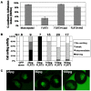
Figure 1. FLRT3 inhibits C-cadherin adhesion activity and induces cell sorting at low expression levels.
A) The effect of FLRT3 expression on blastomere adhesion to C-cadherin coated substrates. Blastomeres were collected from stage 9 embryos that were mock-injected (as control), injected with FLRT3 RNA (160 pg) alone, or co-injected with FLRT3 RNA (160 pg) and C-cadherin RNA (1.5 ng). A portion of the FLRT3 expressing blastomeres was further treated with the Fab fragment of C-cadherin activating antibody, AA5 (1 µg/ml), for 30 min. Nt/No  =  the ratio of the number of blastomeres remaining attached to the C-cadherin substrate after shaking to the number before shaking. B) Dose effects of FLRT3 on its cell sorting activity. Different amounts of FLRT3 RNA, together with NLS-GFP mRNA (200 pg) for a lineage tracer, were injected into one animal blastomere of embryos at the 16-cell stage. The cell sorting activity of FLRT3 was evaluated at stage 13 by observing how much the GFP labeled cells disperse into the uninjected region. C) Representative cell sorting images of embryos injected with 25 pg, 50 pg or 100 pg of FLRT3 RNA.
Figure 2
Figure 2. Effects of FLRT and RND1 knockdowns on PAPC mediated cell adhesion regulation and cell sorting.
A) FLRT morpholinos (FLRT1&3-MOs) and RND1 morpholino (RND1-MO) increase C-cadherin mediated cell adhesion and counteract PAPC-mediated down-regulation of C-cadherin adhesion activity. Embryos were injected as indicated (FL-PAPC or M-PAPC RNA: 1.5 ng, FLRT or RND1 morpholinos: 80 ng) at the 2-4 cell stage. Blastomeres were collected at stage 9 for adhesion assays on C-cadherin coated substrates. B) Comparison of PAPC induced cell sorting versus RND1-MO induced cell sorting by cell reaggregation assay. Either FL-PAPC RNA or RND1-MO was co-injected with NLS-GFP RNA into 2–4 cell stage embryos. At stage 9, dissociated animal cap blastomeres from injected (labeled) embryos were coaggregated with blastomeres from mock injected embryos. The aggregates were bisected and examined under a fluorescence microscope. C) PAPC induces cell sorting in the absence of FLRTs or RND1. FLRT1&3-MOs (80 ng) or RND1-MO (80 ng) were injected into all embryos at the 2–4 cell stage to make all cells deficient in FLRT1&3 or RND1. FL-PAPC RNA (1.5 ng/embryo) and NLS-GFP RNA (or NLS-GFP alone as a control) were injected into one half of the embryos to determine if labeled cells would sort from unlabeled cells. Cell reaggregation assays were performed between labeled (green) and unlabeled cells.
Figure 3
Figure 3. Effects of PAPC expression on the cell sorting and adhesion regulating function of FLRT3.
A) PAPC and FLRT3 cooperate in causing cell sorting. Cell sorting (cell dispersal) assays were performed by injecting 100 pg of FLRT3 RNA, 50 pg of M-PAPC RNA, 100 pg of PAPC-TMC or 100 pg of FL-PAPC RNA separately or in combination into one animal blastomere at the 16 cell stage along with NLS-GFP RNA. B) PAPC expression suppresses the FLRT3-induced disintegration of the blastocoel roof. The indicated RNAs (FLRT3 RNA: 0.4 ng, FL-PAPC or M-PAPC RNA: 1.6 ng) were injected into the animal hemispheres of the 2-4 cell stage embryos, the animal cap explants were excised at stage 9, and the cells of the blastocoel roof were examined and photographed as shown in the diagram. C) Graphical summary of 3 independent experiments examining the PAPC-rescue of the FLRT3 phenotype as shown in B. The results were categorized in terms of severe, mild or no cell adhesion disruption. D) PAPC expression suppresses the downregulation of C-cadherin mediated cell adhesion by FLRT3. Blastomere adhesion assay on C-cadherin coated substrates were performed with cells from embryos injected as in B.
Figure 4
Figure 4. PAPC, FLRT3 and E-cadherin physically interact shown by bead recruitment assay.
A) E-cadherin coated beads specifically recruit PAPC and FLRT3, but not integrin α5 (Intα5), at the surface of transfected A431 cells. Protein A-coupled microbeads were coated with purified human E-cad-EC·Fc and attached to sub-confluent A431 cells that express Xenopus PAPC and/or FLRT3. Double immunofluorescence staining against E-cadherin (red) and PAPC (green), E-cadherin (red) and FLRT3 (green), or E-cadherin (red) and integrin α5 (green). Anti-HLA coated protein A beads were used as negative control. The positions of beads determined by DIC microscopy are marked with arrowheads. White arrowheads indicate beads that did not recruit at least one of the stained proteins, whereas the yellow ones point to those that did recruit E-cadherin, PAPC, and/or FLRT3. B) Quantification of the recruitment of E-cadherin, PAPC and FLRT3 by both anti-HLA control beads and E-cadherin beads as well as the recruitment of integrin α5 by E-cadherin beads.
Figure 5
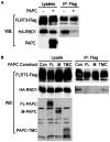
Figure 5. PAPC, FLRT3 and E-cadherin physically interact shown by co-immunoprecipitation (co-IP) assays.
A) PAPC and FLRT3 co-IP with E-cadherin in transfected A431 cells. Parental A431 cells and A431 cells exogenously expressing FLRT3 alone, PAPC alone, or both were grown to confluence in 10 cm petri dishes, lysed in lysis buffer, and IPed with mouse IgG or anti-E-cadherin IgG and analyzed by Western blots against E-cadherin, PAPC, FLRT3 or integrin α5 (as negative control). B) PAPC co-IPs with FLRT3. The same cells as in A) were lysed in lysis buffer, and IPed with rabbit IgG or anti-FLRT3 IgG and analyzed by Western blots for FLRT3 and PAPC.
Figure 6
Figure 6. PAPC expression inhibits the binding of RND1 to FLRT3.
A) Effect of full length PAPC on the binding of RND1 to FLRT3. FLRT3-Flag RNA (0.3 ng) and RND1-HA RNA (0.15 ng) was co-injected into 4-cell stage embryos with or without FL-PAPC RNA (1.5 ng) and extracts of embryos were subject to anti-Flag IP and Western blotting (WB) for the proteins shown. B) The extracellular and transmembrane domains of PAPC inhibit FLRT3 recruitment of RND1. Expression of FL-PAPC and M-PAPC, but not PAPC-TMC, in the Xenopus embryo inhibits FLRT3 recruitment of RND1. Co-IP and Western blot analysis were done as in A.

References

    1. Chen X, Gumbiner BM. Paraxial protocadherin mediates cell sorting and tissue morphogenesis by regulating C-cadherin adhesion activity. J Cell Biol. 2006;174:301–313. - PMC - PubMed
    1. Ogata S, Morokuma J, Hayata T, Kolle G, Niehrs C, et al. TGF-beta signaling-mediated morphogenesis: modulation of cell adhesion via cadherin endocytosis. Genes Dev. 2007;21:1817–1831. - PMC - PubMed
    1. Kim SH, Yamamoto A, Bouwmeester T, Agius E, Robertis EM. The role of paraxial protocadherin in selective adhesion and cell movements of the mesoderm during Xenopus gastrulation. Development. 1998;125:4681–4690. - PubMed
    1. Wunnenberg-Stapleton K, Blitz IL, Hashimoto C, Cho KW. Involvement of the small GTPases XRhoA and XRnd1 in cell adhesion and head formation in early Xenopus development. Development. 1999;126:5339–5351. - PubMed
    1. Brieher WM, Gumbiner BM. Regulation of C-cadherin function during activin induced morphogenesis of Xenopus animal caps. J Cell Biol. 1994;126:519–527. - PMC - PubMed

Publication types

MeSH terms