. 2009 Dec 21;4(12):e8359.
doi: 10.1371/journal.pone.0008359.

Bone marrow osteoblastic niche: a new model to study physiological regulation of megakaryopoiesis

Affiliations

Bone marrow osteoblastic niche: a new model to study physiological regulation of megakaryopoiesis

Isabella Pallotta et al. PLoS One. .

Abstract

Background: The mechanism by which megakaryocytes (Mks) proliferate, differentiate, and release platelets into circulation are not well understood. Growing evidence indicates that a complex regulatory mechanism, involving cellular interactions, composition of the extracellular matrix and physical parameters such as oxygen tension, may contribute to the quiescent or permissive microenvironment related to Mk differentiation and maturation within the bone marrow.

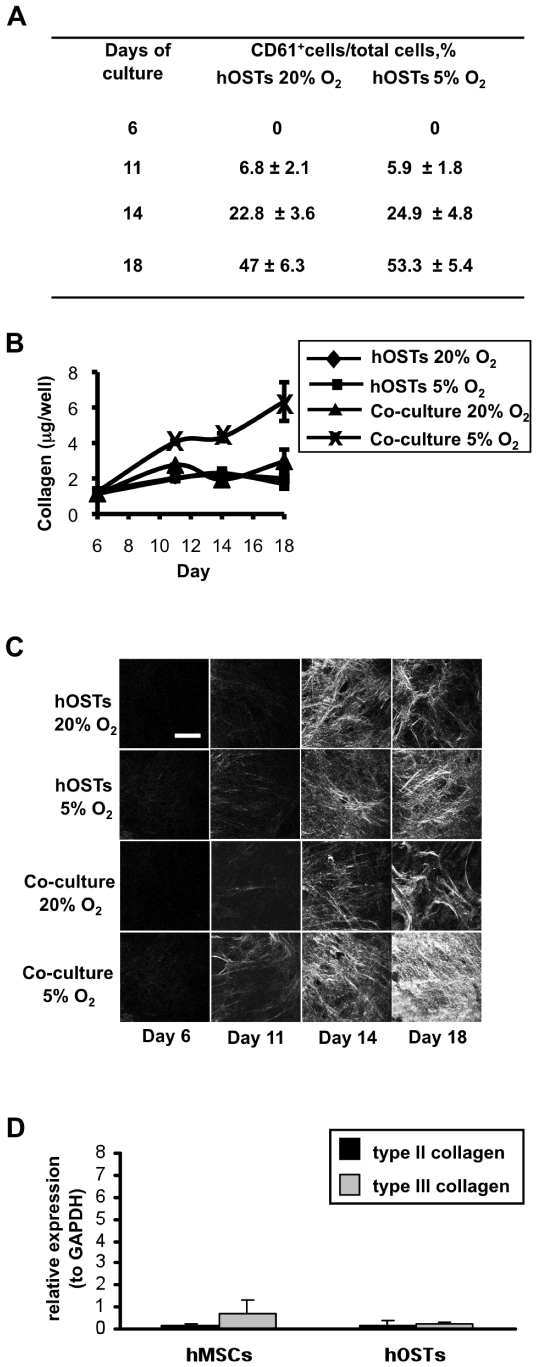
Methodology/principal findings: Differentiating human mesenchymal stem cells (hMSCs) into osteoblasts (hOSTs), we established an in vitro model for the osteoblastic niche. We demonstrated for the first time that the combination of HSCs, Mks and hypoxia sustain and promote bone formation by increasing type I collagen release from hOSTs and enhancing its fibrillar organization, as revealed by second harmonic generation microscopy. Through co-culture, we demonstrated that direct cell-cell contact modulates Mk maturation and differentiation. In particular we showed that low oxygen tension and direct interaction of hematopoietic stem cells (HSCs) with hOSTs inhibits Mk maturation and proplatelet formation (PPF). This regulatory mechanism was dependent on the fibrillar structure of type I collagen released by hOSTs and on the resulting engagement of the alpha2beta1 integrin. In contrast, normoxic conditions and the direct interaction of HSCs with undifferentiated hMSCs promoted Mk maturation and PPF, through a mechanism involving the VCAM-1 pathway.

Conclusions/significance: By combining cellular, physical and biochemical parameters, we mimicked an in vitro model of the osteoblastic niche that provides a physiological quiescent microenvironment where Mk differentiation and PPF are prevented. These findings serve as an important step in developing suitable in vitro systems to use for the study and manipulation of Mk differentiation and maturation in both normal and diseased states.


Conflict of interest statement

Competing Interests: The authors have declared that no competing interests exist.

Figures

Figure 1
Figure 1. Characterization of the osteoblastic niche.
As markers of differentiation we analyzed the expression of bone-related genes, BSP (A), ALP (B) and type I collagen (C) by hOSTs after 6 days of differentiation, as compared to hMSCs, evaluated by real-time RT-PCR. Error bars represent standard deviations. (C) Time course analysis of type I collagen expression by hMSCs and hOSTs at different culture time, evaluated by real-time RT-PCR. (D) Type I collagen protein expression by hMSCs and hOSTs at day 11 of culture. 10 µg of hMSCs and hOSTs lysate and 40 ng of purified type I collagen (C+) were employed in a dot-blot assay. (E) Time course analysis of type I collagen release by hMSCs and hOSTs. The amount of hydroxyproline was measured and then multiplied by a factor of 7.46 to give the total collagen for each well, as described in Materials and Methods. (F) Mineralization of hMSCs (I, II, III) and hOSTs (IV, V, VI)) was evaluated by alizarin red staining, after 6, 11 and 18 days of differentiation (scale bar  = 200 µm). The cultures were performed in normoxic conditions, data are the mean ± SD of the results obtained in three different experiments for each sample. *p<0.05.
Figure 2
Figure 2. Contribution of HSCs, Mks and hypoxia on type I collagen release and organization.
(A) HSC differentiation into Mks was monitored over time and % of CD61+ are reported for each time point. (B) Collagen produced by hOSTs alone or in direct co-culture with HSCs, at different oxygen tensions. The amount of hydroxyproline for each well was evaluated at the indicated time points and converted to collagen, as described in Materials and Methods. Data are the mean ± SD of the results obtained in three different experiments for each sample. (C) Second harmonic generation images of hOSTs at day 6, day 11, day 14, and 18 alone or in direct co-culture with HSCs, performed at O2 20% or at O2 5%. All images were acquired through a 63x (1.2 NA) objective (scale bar  = 30 µm). (D) Expression of type II and III collagen by hOSTs after 6 days of differentiation, as compared to hMSCs, evaluated by real-time RT-PCR.
Figure 3
Figure 3. Role of the osteoblastic niche in megakaryocyte differentiation and proplatelet formation.
(A) HSCs were plated in direct co-culture with hMSCs, or hOSTs or without any feeder layers (control). Adherent cells were counted as described in methods and normalized to relative control suspension cultures. (B) At day 12 of a direct co-culture cells were stained and the percentage of CD61-positive cells was determined upon conventional fluorescence microscopy analysis. Mks were assigned to distinct stages of differentiation as described in Materials and Methods. (C) Representative picture of CD61+ cells at different stage of maturation. (D) Proplatelet-bearing Mks were then evaluated by fluorescence microscopy, after staining with anti-alpha tubulin. Results are expressed as percentage of adherent megakaryocytes forming proplatelets and normalized to relative controls at different oxygen tensions. Data are the mean ± SD of the results obtained in three different experiments for each sample. Statistical analysis by one-way ANOVA followed by Bonferroni's t-test was performed for unpaired observations between hMSCs and hOSTs values. Student t-test was performed for paired observations to relative control. *p<0.05.
Figure 4
Figure 4. Involvement of type I collagen and VCAM-1 in proplatelet formation.
Megakaryocytes, differentiated in suspension cultures for twelve days, were incubated for 30 min with unrelated isotype-matched IgG (control), the monoclonal antibody anti-integrin alpha2, clone P1E6, or anti-integrin alpha4, clone P1H4. Incubated cells, as well as control cells, were allowed to adhere to hMSCs or hOSTs or plated in suspension (control). (A) A reduction of about 30% in Mk adhesion in the presence of the anti-alpha2 antibody on hOSTs was observed, as shown in contrast phase images (scale bar  = 100 µm). (B) The percentage of adherent megakaryocytes extending proplatelets was evaluated after 16 hours. Significant difference in PPF is indicated in comparison to Mks in adhesion to hMSCs or hOSTs without any antibody pre-incubation. (C) Human megakaryocytes were treated with 100 µM blebbistatin or with DMSO for 30 min at 37°C prior to being plated onto hOSTs. PPF was evaluated upon incubation for 16 h. Results are reported as percentage proplatelet-forming cells compared with the corresponding control sample (Mks in suspension), indicated by the dotted line. Data are the mean ± SD of the results obtained in three different experiments for each sample. *p<0.05.
Figure 5
Figure 5. Morphological analysis of proplatelets.
Differential interference contrast (DIC) and immunofluorescence (IF) images of a megakaryocyte extending proplatelets in suspension (A), in adhesion to hMSCs (B), or to hOSTs (C), as revealed by staining for CD61 (scale bar  = 40 µm).

Similar articles

Cited by

References

    1. Patel SR, Hartwig JH, Italiano JE. The biogenesis of platelets from megakaryocyte proplatelets. J Clin Invest. 2005;115:3348–3354. - PMC - PubMed
    1. Schulze H, Shivdasani RA. Mechanisms of thrombopoiesis. J Thromb Haemost. 2005;3:1717–1724. - PubMed
    1. Avecilla ST, Hattori K, Heissig B, Tejada R, Liao F, et al. Chemokine-mediated interaction of hematopoietic progenitors with the bone marrow vascular niche is required for thrombopoiesis. Nat Med. 2004;10:64–71. - PubMed
    1. Larson MK, Watson SP. A product of their environment: do megakaryocytes rely on extracellular cues for proplatelet formation? Platelets. 2006;17:435–440. - PubMed
    1. Leven RM, Tablin F. Extracellular matrix stimulation of guinea pig megakaryocyte proplatelet formation in vitro is mediated through the vitronectin receptor. Exp Hematol. 1992;20:1316–1322. - PubMed

Publication types

MeSH terms