. 2009 Dec 22;4(12):e8390.
doi: 10.1371/journal.pone.0008390.

Expression of the VP2 protein of murine norovirus by a translation termination-reinitiation strategy

Affiliations

Expression of the VP2 protein of murine norovirus by a translation termination-reinitiation strategy

Sawsan Napthine et al. PLoS One. .

Abstract

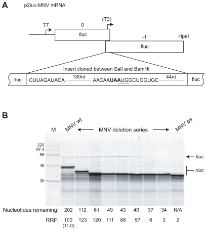
Background: Expression of the minor virion structural protein VP2 of the calicivirus murine norovirus (MNV) is believed to occur by the unusual mechanism of termination codon-dependent reinitiation of translation. In this process, following translation of an upstream open reading frame (ORF) and termination at the stop codon, a proportion of 40S subunits remain associated with the mRNA and reinitiate at the AUG of a downstream ORF, which is typically in close proximity. Consistent with this, the VP2 start codon (AUG) of MNV overlaps the stop codon of the upstream VP1 ORF (UAA) in the pentanucleotide UAAUG.

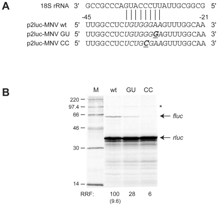
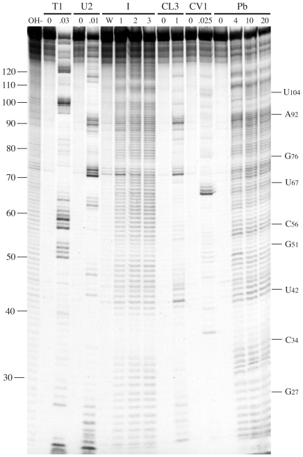
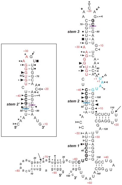
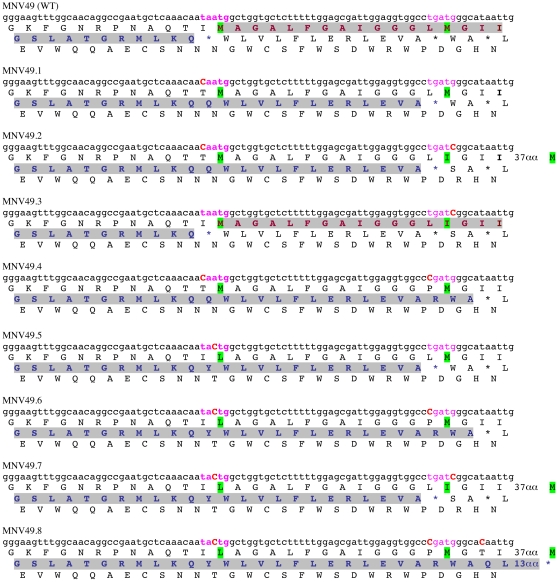
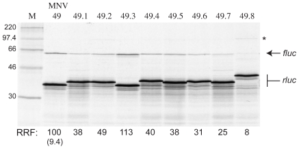
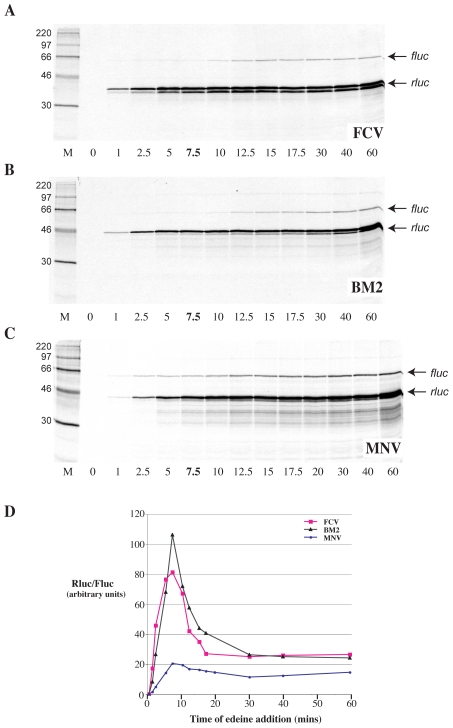
Principal findings: Here, we confirm that MNV VP2 expression is regulated by termination-reinitiation and define the mRNA sequence requirements. Efficient reintiation is dependent upon 43 nt of RNA immediately upstream of the UAAUG site. Chemical and enzymatic probing revealed that the RNA in this region is not highly structured and includes an essential stretch of bases complementary to 18S rRNA helix 26 (Motif 1). The relative position of Motif 1 with respect to the UAAUG site impacts upon the efficiency of the process. Termination-reinitiation in MNV was also found to be relatively insensitive to the initiation inhibitor edeine.

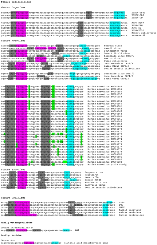
Conclusions: The termination-reinitiation signal of MNV most closely resembles that of influenza BM2. Similar to other viruses that use this strategy, base-pairing between mRNA and rRNA is likely to play a role in tethering the 40S subunit to the mRNA following termination at the VP1 stop codon. Our data also indicate that accurate recognition of the VP2 ORF AUG is not a pre-requisite for efficient reinitiation of translation in this system.


Conflict of interest statement

Competing Interests: The authors have declared that no competing interests exist.

Figures

Figure 1
Figure 1. Minimal sequence requirements for MNV termination-reinitiation.
A) Schematic of the p2luc-MNV reporter mRNA. The termination-reinitiation region (203 nt upstream and 52 nt downstream of the UAA UG motif) was cloned into the SalI and BamHI sites of the p2luc reporter plasmid. HpaI run-off transcripts for in vitro translation were generated using T7 RNA polymerase. The location of the T3 promoter present in the structure mapping construct p2luc-MNV-T3 is indicated. B) Deletion analysis of MNV termination-reinitiation. A series of p2luc-MNV variants were prepared with stepwise, in-frame deletions from the 5′ end of the inserted viral sequence. The wild-type (wt), premature stop (ps) and deletion mutant plasmids were linearised with HpaI and run-off transcripts translated in Flexi® RRL at a final RNA concentration of 50 µg/ml in the presence of [35S]-methionine and 140 mM added KCl. The products were resolved by 12% SDS-PAGE and visualised by autoradiography. The number of nucleotides of viral sequence remaining up to the AUG start codon of the MNV ORF is shown below the gel. The product of the full-length or truncated versions of the rlucVP1 ORF (predicted size of MNVwt is 42 kDa) is marked rluc, and the VP2fluc product (predicted size, 62 kDa) is marked fluc. The MNV ps rluc product is the shortest (predicted size, 33 kDa). RRF denotes the relative reinitiation frequency in comparison to MNVwt (set at 100). The figure in brackets represents the ratio of the intensity of the fluc and rluc products (adjusted for methionine content and expressed as a percentage) for the MNVwt mRNA.
Figure 2
Figure 2. Investigating the role of the MNV 18S rRNA complementary region (Motif 1) in termination-reinitiation.
A) Comparison of part of the sequence of helix 26 of 18S rRNA and the complementary sequence present upstream of the termination-reinitiation site of MNV. Contiguous nucleotides complementary to the 18S rRNA are shown in italics. Putative mRNA-rRNA base pairing is marked, with the mRNA bases numbered relative to the stop codon of rlucVP1. The sequence of the two constructs generated to address the role of the complementary region is also shown, with changes in bold and underlined. B) Plasmids were linearised, transcribed, translated and analysed according to the legend of Figure 1. Lanes are labelled with the last two letters of each reporter plasmid name.
Figure 3
Figure 3. Structure probing of the MNV termination-reinitiation signal.
RNA derived by transcription of p2luc-MNV-T3/BamHI with T3 RNA polymerase was 5′ end-labelled with [γ-33P]-ATP and subjected to limited RNase or chemical cleavage using structure-specific probes. Sites of cleavage were identified by comparison with a ladder of bands created by limited alkaline hydrolysis of the RNA (OH-) and the position of known RNase U2 and T1 cuts, determined empirically. Products were analysed on a 10% acrylamide/7M urea gel containing formamide. Data was also collected from 6% and 15% gels (gels not shown). Enzymatic structure probing was with RNases T1, U2, CL3 and CV1. Uniquely cleaved nucleotides were identified by their absence in untreated control lanes (0). The number of units of enzyme added to each reaction is indicated. Chemical structure probing was with imidazole (I, hours) or lead acetate (Pb; mM concentration in reaction). The water lane (W) represents RNA which was dissolved in water, incubated for four hours and processed in parallel to the imidazole-treated sample. The sequence of the probed RNA and the inferred secondary structure is shown in Figure 4.
Figure 4
Figure 4. Summary of the MNV structure probing results.
The sensitivity of bases in the MNV termination-reinitiation region to the various probes is shown for an mfold prediction (see text). The first base of the transcript is numbered 1. The bases are also numbered (in red) with respect to the VP1 stop codon (with the U of the UAA codon numbered +1, the preceding base numbered -1). The reactivies of the T1 (black triangle), U2 (asterisk), CL3 (open triangle) and CV1 (black square) probes are marked. The size of the symbols is approximately proportional to the intensity of cleavage at that site. Lead and imidazole cleavages are not marked, but bases resistant to cleavage by both reagents are shown in bold/outline font. The two large arrows show the boundaries beyond which no structure mapping information was obtained. The stretch of bases in red indicate the 18S rRNA complementary region. Bases that form the stop-start overlap are in blue. The blue line indicates the start of the minimal essential region required for efficient termination-reinitiation. The purple line indicates the likely location of the 5′-edge of a ribosome poised at the termination codon (UAA, in blue). Bases in lower case are of vector origin. The mfold shown in the box shows part of an alternative pairing possibility in which the 5′ arm of stem 2 pairs with a different region (to give stem 2′; see text).
Figure 5
Figure 5. The effect of moving the stop codon of the termination-reinitiation window further downstream on the mRNA.
A series of plasmid constructs were prepared, based on MNV.49 (a fully functional, truncated version of pMNVwt [see Figure 1] which acts as the “wild-type” reference construct [WT] in these experiments), in which the stop and start codons of the termination-reinitiation signal were altered. The figure shows the primary sequence and three-frame translation of the relevant region of the mRNA encoded by each construct. The natural stop-start motif is shown in pink and emboldened text, the downstream fortuitous stop-start motif in pink. Mutations within the mRNA sequence are highlighted by uppercase, red emboldened characters. The upstream rlucVP1 ORF is highlighted in grey, as is the downstream VP2fluc ORF where this is known. Likely key methionines (start codons) or their replacement amino acid are highlighted in green.
Figure 6
Figure 6. Effect of moving the stop codon of the termination-reinitiation window further downstream on the mRNA.
The plasmid constructs of Figure 5 were linearised with HpaI and run-off transcripts translated and analysed as decribed in the legend to Figure 1. The product of the full-length or truncated versions of the rlucVP1 ORF is marked rluc, and the VP2fluc product (predicted size, 62 kDa) is marked fluc. The longer product observed in the 49.8 translation is asterisked.
Figure 7
Figure 7. Effect of edeine on termination-reinitiation.
Reporter mRNAs containing the termination reinitiation signals of FCV (panel A), BM2 (panel B) and MNV (Panel C) were translated in Flexi® RRL at a final RNA concentration of 50 µg/ml in the presence of [35S]-methionine and 140 mM added KCl. At the indicated time points (min), an aliquot was removed, edeine added to 5 µM, and the sample reincubated for a total of 60 min. The translation products were resolved by SDS-PAGE on 12% gels and visualised by autoradiography. Identical experiments were performed in which cycloheximide replaced edeine (data not shown). In the cycloheximide experiments, it was found that in all cases, no termination-reinitiation product was evident until the 7.5 min time point, when only a trace was visible. The 7.5 min time point in the edeine gels is emboldened to reflect this. The relative levels of the rluc and fluc bands was determined by densitometry and in Panel D, the Rluc/Fluc ratio is plotted against the time of edeine addition for the three mRNAs.
Figure 8
Figure 8. Comparison of caliciviral termination-reinitiation signals and 5′ flanking regions.
The termination-reinitiation signal of influenza BM2 is also shown, as is a putative signal in the cellular gene glutamic acid decarboxylase . Confirmed and potential Motif 1 sequences are highlighted in pink and the stop-start window in blue. Potential base-pairing interactions flanking Motif 1 are indicated in grey (or underlined in the case of the glutamic acid decarboxylase gene). Within the murine noroviruses, in reference to EU004666, base changes are highlighted in green. Abbreviations used: EBHSV, European brown hare syndrome virus; RHDV, rabbit hemorrhagic disease virus; VESV, vesicular exanthema of swine virus; FCV, feline calicivirus; SMSV, San Miguel sealion virus.

Similar articles

Cited by

References

    1. Pestova TV, Kolupaeva VG, Lomakin IB, Pilipenko EV, Shatsky IN, et al. Molecular mechanisms of translation initiation in eukaryotes. Proc Natl Acad Sci U S A. 2001;98:7029–7036. - PMC - PubMed
    1. Kozak M. Translation of insulin-related polypeptides from messenger RNAs with tandemly reiterated copies of the ribosome binding site. Cell. 1983;34:971–978. - PubMed
    1. Gale M, Jr, Tan SL, Katze MG. Translational control of viral gene expression in eukaryotes. Microbiol Mol Biol Rev. 2000;64:239–280. - PMC - PubMed
    1. Pavlakis GN, Felber BK. Regulation of expression of human immunodeficiency virus. New Biol. 1990;2:20–31. - PubMed
    1. Wilson JE, Powell MJ, Hoover SE, Sarnow P. Naturally occurring dicistronic cricket paralysis virus RNA is regulated by two internal ribosome entry sites. Mol Cell Biol. 2000;20:4990–4999. - PMC - PubMed

Publication types

MeSH terms