Review
. 2009 Nov-Dec;17(6):544-54.
doi: 10.1590/s1678-77572009000600002.

Cytotoxicity and biocompatibility of direct and indirect pulp capping materials

Affiliations
Review

Cytotoxicity and biocompatibility of direct and indirect pulp capping materials

Karin Cristina da Silva Modena et al. J Appl Oral Sci. 2009 Nov-Dec.

Abstract

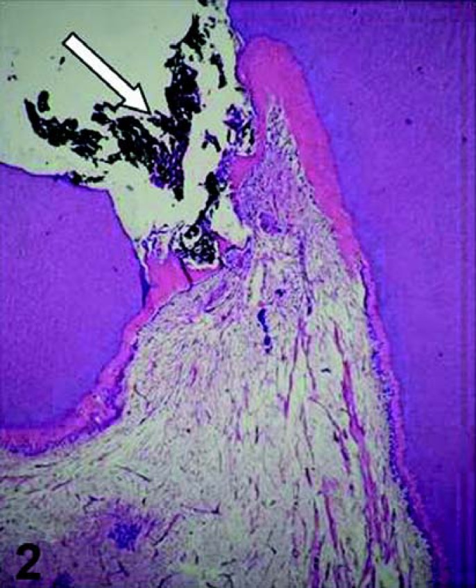
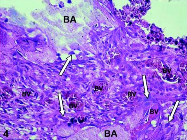
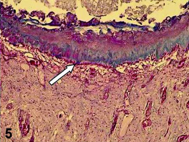
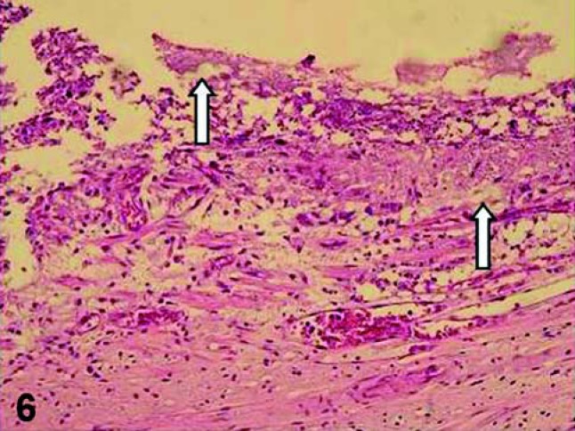
There are several studies about the cytotoxic effects of dental materials in contact with the pulp tissue, such as calcium hydroxide (CH), adhesive systems, resin composite and glass ionomer cements. The aim of this review article was to summarize and discuss the cytotoxicity and biocompatibility of materials used for protection of the dentin-pulp complex, some components of resin composites and adhesive systems when placed in direct or indirect contact with the pulp tissue. A large number of dental materials present cytotoxic effects when applied close or directly to the pulp, and the only material that seems to stimulate early pulp repair and dentin hard tissue barrier formation is CH.


Figures

FIGURE 1
FIGURE 1. General view of the dentin-pulp complex. Below the tubular dentin (D) there is the predentin (Pd), which is underlined by the odontoblast layer (Od). Observe the cellfree zone of Weil – horizontal arrow) and the cell-rich zone (vertical arrows). HE, ×64
FIGURE 2
FIGURE 2. Pulp exposure capped with calcium hydroxide (arrow). Observe that 30 days after the pulp therapy, a partial hard tissue barrier was formed adjacent to the capping agent. HE, ×32
FIGURE 3
FIGURE 3. Sixty days after applying calcium hydroxide on the pulp tissue, a complete hard tissue barrier (HB) is formed. Note the tunnel defect (horizontal arrow) and cellular inclusions (vertical arrows) within the hard tissue barrier, which is underlined by a new layer of odontoblast-like cells. HE, ×125
FIGURE 4
FIGURE 4. Exposed human pulp tissue capped with an adhesive system. Sixty days after the capping procedure the tooth was extracted and processed for histological evaluation of the pulp tissue. Note the presence of bonding agent (BA) on the pulp exposure site and inside of the connective tissue (BA – arrow). Intense chronic inflammatoty reaction mediated by macrophages and a number of dilated and congested blood vessles (BV) is observed. HE, ×320
FIGURE 5
FIGURE 5. Pulp capped with a dental material that present high pH (MTA). Sixty days after the pulp therapy, a defined hard tissue barrier was formed, which was underlined by a new layer of odontoblast-like cells (arrow). Masson's Trichrome, ×86
FIGURE 6
FIGURE 6. Pulp exposure capped with an adhesive system. Note that no hard tissue barrier was formed 60 days after the pulp therapy. Fragments of resinous material (arrows) are observed displaced to the pulp exposure site, which exhibits persistent inflammatory response. HE, ×125
FIGURE 7
FIGURE 7. Deep cavity was prepared in human premolar. In this figure it is shown the pulp tissue related to the cavity floor in which total etch was applied and an adhesive system and composite resin were used for cavity restoration. Sixty days after the clinical procedure the tooth was extracted and processed for histological evaluation. Note the intense inflammatory response as well as the zone of inner dentinal resorption (arrow). HE, ×160

References

    1. Abbas A, Lichtman A. Cellular and molecular immunology. In: Abbas A, Lichtman A, editors. Cytokines. 5 ed. Philadelphia: Saunders; 2003. pp. 243–274.
    1. Aeinehchi M, Eslami B, Ghanbariha M, Saffar AS. Mineral trioxide aggregate (MTA) and calcium hydroxide as pulp-capping agents in human teeth: a preliminary report. Int Endod J. 2002;36:225–231. - PubMed
    1. Akimoto N, Momoi Y, Kohno A, Suzuki S, Otsuki M, Suzuki S, et al. Biocompatibility of Clearfil Liner Bond 2 and Clearfil AP-X system on nonexposed and exposed primate teeth. Quintessence Int. 1998;29(3):177–188. - PubMed
    1. Aranha AMF, Giro EMA, Souza PPC, Hebling J, Costa CAS. Effect of curing regime on the cytotoxicity of resin-modified glass-ionomer lining cements applied to an odontoblast-cell line. Dent Mater. 2006;22:864–869. - PubMed
    1. Barkhordar R, Ghani Q, Russell T, Hussain M. Interleukin-1beta activity and collagen synthesis in human dental pulp fibroblasts. J Endod. 2002;28(3):157–159. - PubMed