

Assessment of canal walls after biomechanical preparation of root canals instrumented with Protaper Universal rotary system
- PMID: 20027432
- PMCID: PMC4327519
- DOI: 10.1590/s1678-77572009000600010
Assessment of canal walls after biomechanical preparation of root canals instrumented with Protaper Universal rotary system
Abstract
Objective: The aim of this study was to examine the instrumented walls of root canals prepared with the ProTaper Universal rotary system.
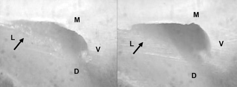
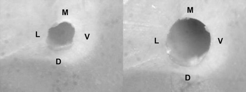
Material and methods: Twenty mesiobuccal canals of human first mandibular molars were divided into 2 groups of 10 specimens each and embedded in a muffle system. The root canals were transversely sectioned 3 mm short of the apex before preparation and remounted in their molds. All root canals were prepared with ProTaper Universal rotary system or with Nitiflex files. The pre and postoperative images of the apical thirds viewed with a stereoscopic magnifier (X45) were captured digitally for further analysis. Data were analyzed statistically by Fisher's exact test and Chi-square test at 5% significance level.
Results: The differences observed between the instrumented and the noninstrumented walls were not statistically significant (p<0.05).
Conclusions: The Nitiflex files and the ProTaper Universal rotary system failed to instrument all the root canal walls.
Figures
Similar articles
-
Evaluation of the centreing ability of the ProTaper Universal rotary system in curved roots in comparison to Nitiflex files.Aust Endod J. 2009 Dec;35(3):174-9. doi: 10.1111/j.1747-4477.2009.00168.x. Aust Endod J. 2009. PMID: 19961458 Clinical Trial.
-
Instrument separation analysis of multi-used ProTaper Universal rotary system during root canal therapy.J Endod. 2011 Jun;37(6):758-63. doi: 10.1016/j.joen.2011.02.021. Epub 2011 Apr 2. J Endod. 2011. PMID: 21787484
-
Root canal preparation of mandibular molars with 3 nickel-titanium rotary instruments: a micro-computed tomographic study.J Endod. 2014 Nov;40(11):1860-4. doi: 10.1016/j.joen.2014.06.023. Epub 2014 Sep 6. J Endod. 2014. PMID: 25205262
-
ProTaper rotary instrument fracture during root canal preparation: a comparison between rotary and hybrid techniques.Oral Health Dent Manag. 2013 Mar;12(1):50-5. Oral Health Dent Manag. 2013. PMID: 23474581 Clinical Trial.
-
Root canal shaping using rotary nickel-titanium files in preclinical dental education in Turkey.J Dent Educ. 2012 Apr;76(4):509-13. J Dent Educ. 2012. PMID: 22473564
Cited by
-
Apical dye leakage of two single-cone root canal core materials (hydrophilic core material and gutta-percha) sealed by different types of endodontic sealers: An in vitro study.J Conserv Dent. 2018 Mar-Apr;21(2):147-152. doi: 10.4103/JCD.JCD_154_17. J Conserv Dent. 2018. PMID: 29674815 Free PMC article.
-
Apical transportation associated with ProTaper Universal F1, F2 and F3 instruments in curved canals prepared by undergraduate students.J Appl Oral Sci. 2014 Apr;22(2):98-102. doi: 10.1590/1678-775720130464. J Appl Oral Sci. 2014. PMID: 24676579 Free PMC article.
-
In vitro comparison of bioceramic and silicone-based root canal sealers with different obturation technique.J Conserv Dent Endod. 2025 Jun;28(6):527-531. doi: 10.4103/JCDE.JCDE_175_25. Epub 2025 Jun 2. J Conserv Dent Endod. 2025. PMID: 40546863 Free PMC article.
-
Assessment of Biomechanical Preparation Influence on Various Root Canal Curvatures.Int J Clin Pediatr Dent. 2024 Feb;17(2):130-135. doi: 10.5005/jp-journals-10005-2760. Int J Clin Pediatr Dent. 2024. PMID: 39184879 Free PMC article.
-
Comparison of debris extruded apically and working time used by ProTaper Universal rotary and ProTaper retreatment system during gutta-percha removal.J Appl Oral Sci. 2010 Dec;18(6):542-5. doi: 10.1590/s1678-77572010000600002. J Appl Oral Sci. 2010. PMID: 21308282 Free PMC article.
References
-
- Aguiar CM, Câmara AC. Radiological evaluation of the morphological changes of root canals shaped with ProTaper™ for hand use and the ProTaper™ and RaCe™ rotary instruments. Aust Endod J. 2008;34:115–119. - PubMed
-
- Aguiar CM, Mendes DA, Câmara AC, Figueiredo JAP. Evaluation of the centreing ability of the ProTaper Universal™ rotary system in curved roots in comparison to Nitiflex™ files. Aust Endod J. 2009. [cited 2009 Nov 4]. serial on the internet. In press. Available from: http://dx.doi.org/10.1111/j.1747-4477.2009.00168.x. - DOI - PubMed
-
- Calberson FL, Deroose CAJ, Hommez GM, De Moor RJ. Shaping ability of ProTaper nickel-titanium files in simulated resin root canals. Int Endod J. 2004;37:613–623. - PubMed
-
- Câmara AC, Aguiar CM, Figueiredo JAP. Assessment of the deviation after biomechanical preparation of the coronal, middle and apical thirds of root canals instrumented with three Hero Rotary Systems. J Endod. 2007;33:1460–1463. - PubMed
Publication types
MeSH terms
Substances
LinkOut - more resources
Full Text Sources
Miscellaneous
