

Intraoperative near-infrared fluorescence imaging in perforator flap reconstruction: current research and early clinical experience
- PMID: 20027541
- PMCID: PMC3100538
- DOI: 10.1055/s-0029-1244805
Intraoperative near-infrared fluorescence imaging in perforator flap reconstruction: current research and early clinical experience
Abstract
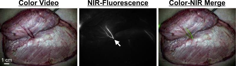
Despite recent advances in perforator flap reconstruction, there can be significant variability in vessel size and location. Although preoperative evaluation may provide valuable information, real-time intraoperative methods have the potential to provide the greatest benefit. Our laboratory has developed the Fluorescence-Assisted Resection and Exploration (FLARE) near-infrared (NIR) fluorescence imaging system for intraoperative visualization of details of the underlying vasculature. The FLARE system uses indocyanine green, a safe and reliable NIR fluorophore already FDA-approved for other indications. The system has been optimized in large-animal models for the identification of perforator size, location, and perfusion and has also been translated to the clinic for use during breast reconstruction after mastectomy. In this article, we review our preclinical and clinical data, as well as literature describing the use of similar NIR fluorescence imaging systems in plastic and reconstructive surgery.
Figures


Similar articles
-
The FLARE intraoperative near-infrared fluorescence imaging system: a first-in-human clinical trial in perforator flap breast reconstruction.Plast Reconstr Surg. 2010 Nov;126(5):1472-1481. doi: 10.1097/PRS.0b013e3181f059c7. Plast Reconstr Surg. 2010. PMID: 21042103 Free PMC article. Clinical Trial.
-
Anatomic imaging of abdominal perforator flaps without ionizing radiation: seeing is believing with magnetic resonance imaging angiography.J Reconstr Microsurg. 2010 Jan;26(1):37-44. doi: 10.1055/s-0029-1220862. Epub 2009 May 18. J Reconstr Microsurg. 2010. PMID: 19452440
-
The value of the multidetector row computed tomography for the preoperative planning of deep inferior epigastric artery perforator flap: our experience in 162 cases.Ann Plast Surg. 2008 Jan;60(1):29-36. doi: 10.1097/SAP.0b013e31805003c2. Ann Plast Surg. 2008. PMID: 18281792
-
Reviewing the vascular supply of the anterior abdominal wall: redefining anatomy for increasingly refined surgery.Clin Anat. 2008 Mar;21(2):89-98. doi: 10.1002/ca.20585. Clin Anat. 2008. PMID: 18189276 Review.
-
Breast reconstruction with the deep inferior epigastric perforator flap: history and an update on current technique.J Plast Reconstr Aesthet Surg. 2006;59(6):571-9. doi: 10.1016/j.bjps.2006.01.004. Epub 2006 Mar 20. J Plast Reconstr Aesthet Surg. 2006. PMID: 16716950 Review.
Cited by
-
Topical application of activity-based probes for visualization of brain tumor tissue.PLoS One. 2012;7(3):e33060. doi: 10.1371/journal.pone.0033060. Epub 2012 Mar 13. PLoS One. 2012. PMID: 22427947 Free PMC article.
-
The clinical use of indocyanine green as a near-infrared fluorescent contrast agent for image-guided oncologic surgery.J Surg Oncol. 2011 Sep 1;104(3):323-32. doi: 10.1002/jso.21943. Epub 2011 Apr 14. J Surg Oncol. 2011. PMID: 21495033 Free PMC article. Review.
-
Predicting the survival of experimental ischaemic small bowel using intraoperative near-infrared fluorescence angiography.Br J Surg. 2011 Dec;98(12):1725-34. doi: 10.1002/bjs.7698. Epub 2011 Sep 27. Br J Surg. 2011. PMID: 21953541 Free PMC article.
-
A wireless infrared thermometry device for postoperative flap monitoring: Proof of concept in a porcine flap model.Int Wound J. 2023 Aug;20(6):1839-1848. doi: 10.1111/iwj.14034. Epub 2022 Dec 19. Int Wound J. 2023. PMID: 36535065 Free PMC article.
-
A review of indocyanine green fluorescent imaging in surgery.Int J Biomed Imaging. 2012;2012:940585. doi: 10.1155/2012/940585. Epub 2012 Apr 22. Int J Biomed Imaging. 2012. PMID: 22577366 Free PMC article.
References
-
- De Grand AM, Frangioni JV. An operational near-infrared fluorescence imaging system prototype for large animal surgery. Technol Cancer Res Treat. 2003;2:553–562. - PubMed
Publication types
MeSH terms
Grants and funding
LinkOut - more resources
Full Text Sources
Other Literature Sources
Medical
Miscellaneous
