

Early identification of aortic valve sclerosis using iron oxide enhanced MRI
- PMID: 20027578
- PMCID: PMC5638659
- DOI: 10.1002/jmri.22008
Early identification of aortic valve sclerosis using iron oxide enhanced MRI
Abstract
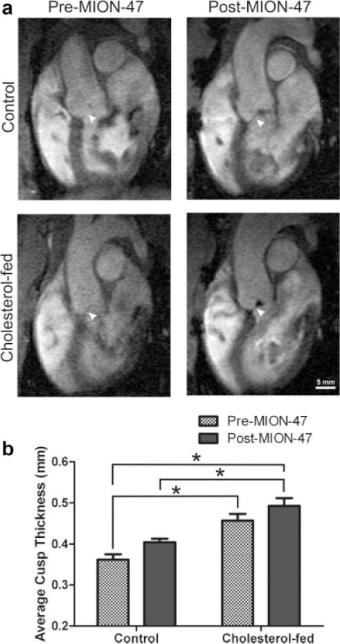
Purpose: To test the ability of MION-47 enhanced MRI to identify tissue macrophage infiltration in a rabbit model of aortic valve sclerosis (AVS).
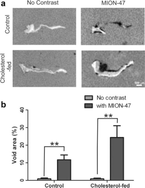
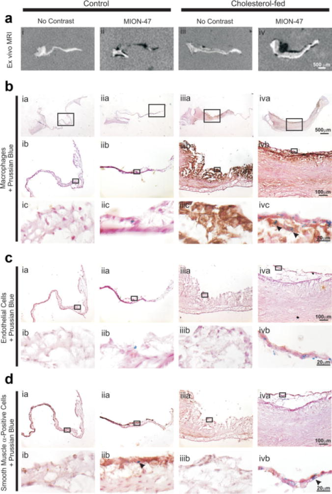
Materials and methods: The aortic valves of control and cholesterol-fed New Zealand White rabbits were imaged in vivo pre- and 48 h post-intravenous administration of MION-47 using a 1.5 Tesla (T) MR clinical scanner and a CINE fSPGR sequence. MION-47 aortic valve cusps were imaged ex vivo on a 3.0T whole-body MR system with a custom gradient insert coil and a three-dimensional (3D) FIESTA sequence and compared with aortic valve cusps from control and cholesterol-fed contrast-free rabbits. Histopathological analysis was performed to determine the site of iron oxide uptake.
Results: MION-47 enhanced the visibility of both control and cholesterol-fed rabbit valves in in vivo images. Ex vivo image analysis confirmed the presence of significant signal voids in contrast-administered aortic valves. Signal voids were not observed in contrast-free valve cusps. In MION-47 administered rabbits, histopathological analysis revealed iron staining not only in fibrosal macrophages of cholesterol-fed valves but also in myofibroblasts from control and cholesterol-fed valves.
Conclusion: Although iron oxide labeling of macrophage infiltration in AVS has the potential to detect the disease process early, a macrophage-specific iron compound rather than passive targeting may be required.
(c) 2009 Wiley-Liss, Inc.
Figures
References
-
- Stewart MD, Siscovick MD, Lind MS, et al. Clinical factors associated with calcific aortic valve disease. J Am Coll Cardiol. 1997;29:630–634. - PubMed
-
- O’Brien KD. Pathogenesis of calcific aortic valve disease: a disease process comes of age (and a good deal more) Arterioscler Thromb Vasc Biol. 2006;26:1721–1728. - PubMed
-
- Lindroos M, Kupari M, Heikkila J, Tilvis R. Prevalence of aortic valve abnormalities in the elderly: an echocardiographic study of a random population sample. J Am Coll Cardiol. 1993;21:1220–1225. - PubMed
-
- Yeo K, Low R. Aortic stenosis: assessment of the patient at risk. J Magn Reson Imaging. 2007;20:509–516. - PubMed
-
- Otto C, Kuusisto J, Reichenbach D, Gown A, O’Brien KD. Characterization of the early lesion of ’degenerative’ valvular aortic stenosis. Histological and immunohistochemical studies. Circulation. 1994;90:844–853. - PubMed
Publication types
MeSH terms
Substances
Grants and funding
LinkOut - more resources
Full Text Sources
Medical
