

Carcinoma of the papilla of Vater following treatment of pancreaticobiliary maljunction
- PMID: 20027689
- PMCID: PMC2797673
- DOI: 10.3748/wjg.15.6126
Carcinoma of the papilla of Vater following treatment of pancreaticobiliary maljunction
Abstract
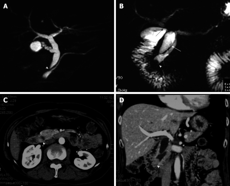
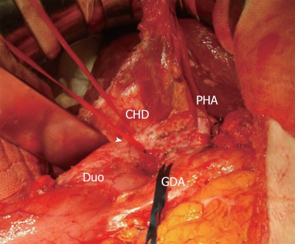
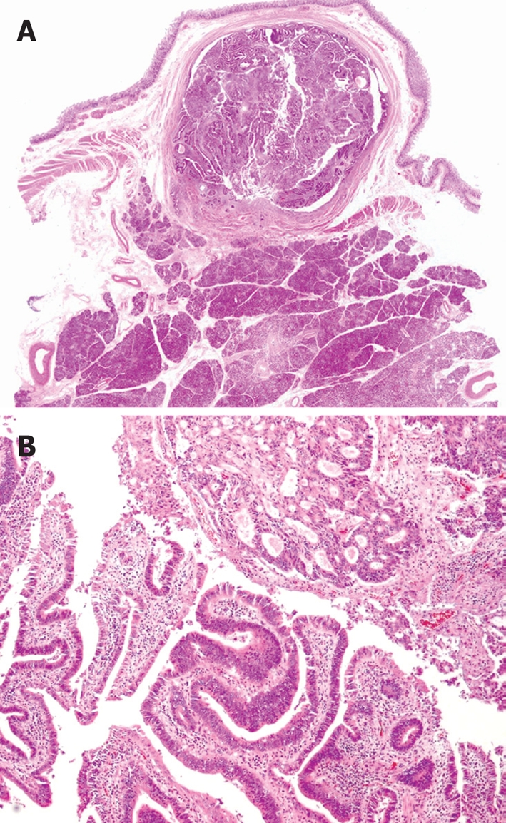
Pancreaticobiliary maljunction (PBM) is frequently associated with biliary cancer due to reflux of pancreatic enzymes into the choledochus, and even after surgery to correct the PBM such patients still have a risk of residual bile duct cancer. Here, we report the case of a 59-year-old female with carcinoma of the papilla of Vater which developed 2.5 years after choledochoduodenostomy for PBM. During the postoperative follow-up period, computed tomography obtained 2 years after the first operation demonstrated a tumor in the distal end of the choledochus, although she did not have jaundice and laboratory tests showed no abnormalities caused by the previous operation. As a result, carcinoma of the papilla of Vater was diagnosed at an early stage, followed by surgical cure. For early detection of periampullary cancer in patients undergoing surgery for PBM, careful long-term follow-up is needed.
Figures
Similar articles
-
Carcinoma of the Papilla of Vater after Diversion Operation for Pancreaticobiliary Maljunction.Case Rep Gastroenterol. 2017 May 17;11(2):265-270. doi: 10.1159/000462967. eCollection 2017 May-Aug. Case Rep Gastroenterol. 2017. PMID: 28626371 Free PMC article.
-
A case report of papilla Vater carcinoma showing positive expression of thymidine phosphorylase.Hepatogastroenterology. 2004 Mar-Apr;51(56):375-7. Hepatogastroenterology. 2004. PMID: 15086163
-
Risk of bile duct carcinogenesis after excision of extrahepatic bile ducts in pancreaticobiliary maljunction.Surgery. 1999 Nov;126(5):939-44. doi: 10.1016/s0039-6060(99)70036-x. Surgery. 1999. PMID: 10568195
-
Pancreaticobiliary maljunction and carcinogenesis to biliary and pancreatic malignancy.Langenbecks Arch Surg. 2009 Jan;394(1):159-69. doi: 10.1007/s00423-008-0336-0. Epub 2008 May 24. Langenbecks Arch Surg. 2009. PMID: 18500533 Review.
-
Recent advances in pancreaticobiliary maljunction.J Hepatobiliary Pancreat Surg. 2002;9(1):45-54. doi: 10.1007/s005340200004. J Hepatobiliary Pancreat Surg. 2002. PMID: 12021897 Review.
Cited by
-
A case report of carcinoma of the papilla of Vater associated with a hyperplasia-dysplasia-carcinoma sequence by pancreaticobiliary maljunction.World J Surg Oncol. 2024 Feb 22;22(1):63. doi: 10.1186/s12957-024-03347-z. World J Surg Oncol. 2024. PMID: 38389074 Free PMC article.
-
Application of imaging techniques in pancreaticobiliary maljunction.World J Clin Cases. 2022 Aug 6;10(22):7642-7652. doi: 10.12998/wjcc.v10.i22.7642. World J Clin Cases. 2022. PMID: 36158479 Free PMC article. Review.
-
Carcinoma of the Papilla of Vater after Diversion Operation for Pancreaticobiliary Maljunction.Case Rep Gastroenterol. 2017 May 17;11(2):265-270. doi: 10.1159/000462967. eCollection 2017 May-Aug. Case Rep Gastroenterol. 2017. PMID: 28626371 Free PMC article.
-
Endoscopic retrograde cholangiopancreatography in children with symptomatic pancreaticobiliary maljunction: A retrospective multicenter study.World J Gastroenterol. 2019 Oct 28;25(40):6107-6115. doi: 10.3748/wjg.v25.i40.6107. World J Gastroenterol. 2019. PMID: 31686766 Free PMC article.
References
-
- The Japanese Study Group on Pancreaticobiliary Maljunction (JSPBM), The Committee of JSPBM for Diagnostic Criteria. Diagnostic criteria of pancreaticobiliary maljunction. J Hepatobiliary Pancreat Surg. 1994;1:219–221.
-
- Tashiro S, Imaizumi T, Ohkawa H, Okada A, Katoh T, Kawaharada Y, Shimada H, Takamatsu H, Miyake H, Todani T. Pancreaticobiliary maljunction: retrospective and nationwide survey in Japan. J Hepatobiliary Pancreat Surg. 2003;10:345–351. - PubMed
-
- Todani T, Watanabe Y, Urushihara N, Morotomi Y, Maeba T. Choledochal cyst, pancreatobiliary malunion, and cancer. J Hepatobiliary Pancreat Surg. 1994;1:247–251.
-
- Ishibashi T, Kasahara K, Yasuda Y, Nagai H, Makino S, Kanazawa K. Malignant change in the biliary tract after excision of choledochal cyst. Br J Surg. 1997;84:1687–1691. - PubMed
-
- Kobayashi S, Asano T, Yamasaki M, Kenmochi T, Nakagohri T, Ochiai T. Risk of bile duct carcinogenesis after excision of extrahepatic bile ducts in pancreaticobiliary maljunction. Surgery. 1999;126:939–944. - PubMed
Publication types
MeSH terms
LinkOut - more resources
Full Text Sources
Medical
Research Materials
