Comparative Study
. 2010 Feb 9;49(5):872-81.
doi: 10.1021/bi901751b.

The ability of rodent islet amyloid polypeptide to inhibit amyloid formation by human islet amyloid polypeptide has important implications for the mechanism of amyloid formation and the design of inhibitors

Affiliations
Comparative Study

The ability of rodent islet amyloid polypeptide to inhibit amyloid formation by human islet amyloid polypeptide has important implications for the mechanism of amyloid formation and the design of inhibitors

Ping Cao et al. Biochemistry. .

Abstract

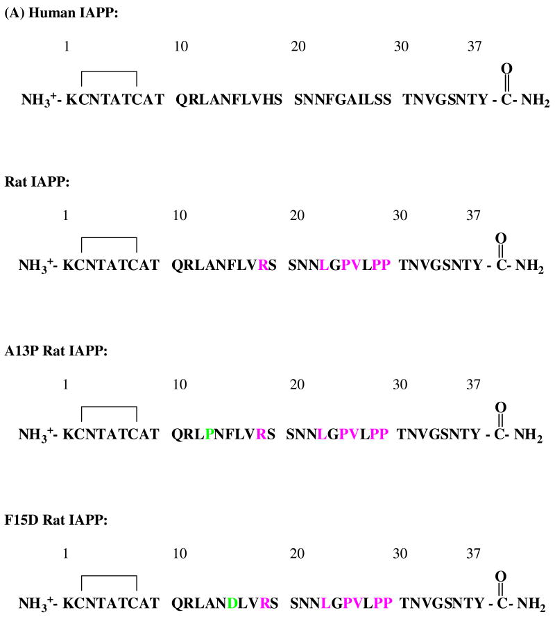
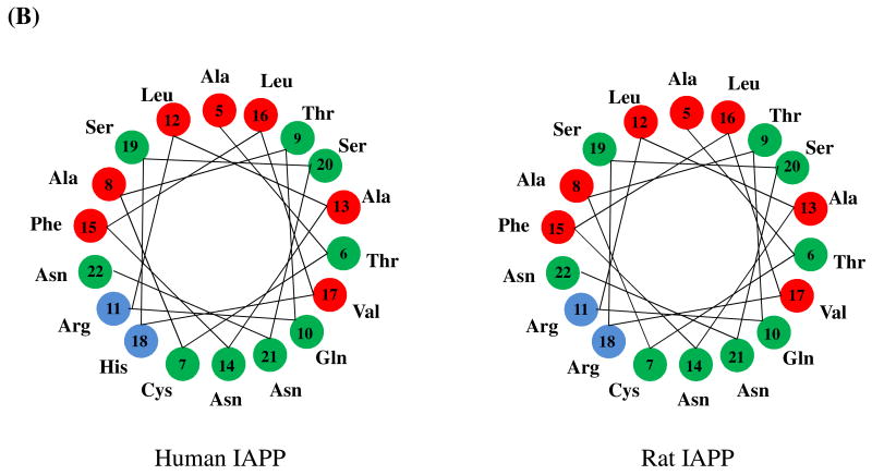
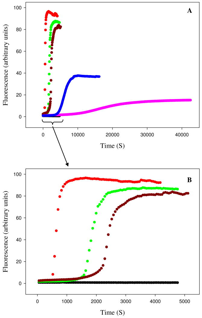
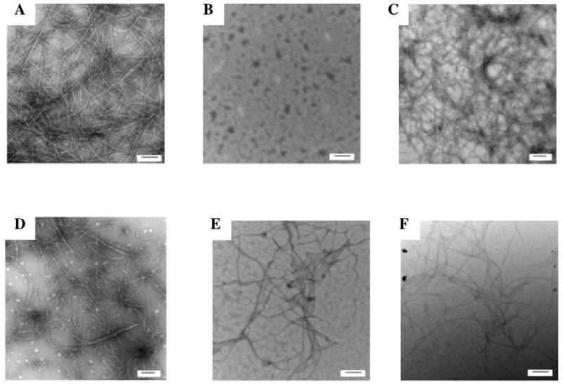
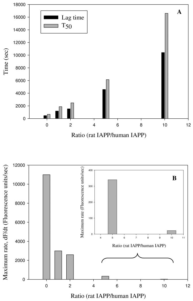
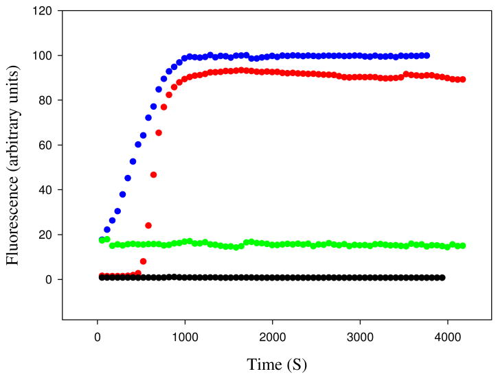
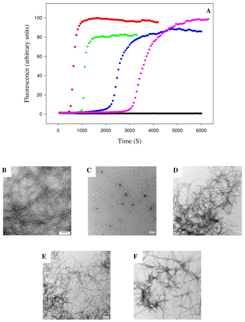
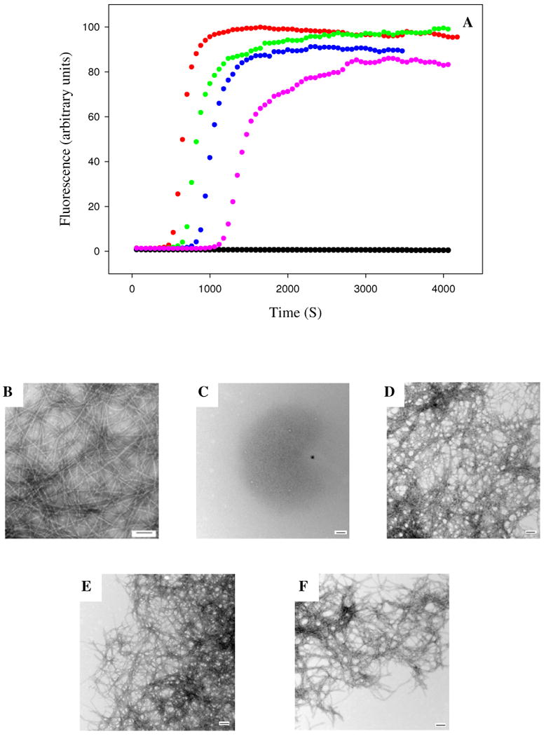
Islet amyloid polypeptide (IAPP) is a 37-residue polypeptide hormone that is responsible for islet amyloid formation in type II diabetes. Human IAPP is extremely amyloidogenic, while rat IAPP and mouse IAPP do not form amyloid in vitro or in vivo. Rat IAPP and mouse IAPP have identical primary sequences, but differ from the human polypeptide at six positions, five of which are localized between residues 20 and 29. The ability of rat IAPP to inhibit amyloid formation by human IAPP was tested, and the rat peptide was found to be an effective inhibitor. Thioflavin-T fluorescence-monitored kinetic experiments, transmission electron microscopy, and circular dichroism showed that rat IAPP lengthened the lag phase for amyloid formation by human IAPP, slowed the growth rate, reduced the amount of amyloid fibrils produced in a dose-dependent manner, and altered the morphology of the fibrils. The inhibition of human IAPP amyloid formation by rat IAPP can be rationalized by a model that postulates formation of an early helical intermediate during amyloid formation where the helical region is localized to the N-terminal region of IAPP. The model predicts that proline mutations in the putative helical region should lead to ineffective inhibitors as should mutations that alter the peptide-peptide interaction interface. We confirmed this by testing the ability of A13P and F15D point mutants of rat IAPP to inhibit amyloid formation by human IAPP. Both these mutants were noticeably less effective inhibitors than wild-type rat IAPP. The implications for inhibitor design are discussed.


Figures

Figure 1
Figure 1
Comparison of human and rat IAPP. (A) Primary sequence of human IAPP, rat IAPP and the A13P and F15D mutants of rat IAPP. All peptides have a disulfide bridge between Cys-2 and Cys-7 and have an amidated C-terminus. Residues which differ from the human peptide are colored in pink. Residues in the mutants which differ from wild type rat IAPP are colored in light green. (B) Helical wheel representation of residues 5 to 22 of human and rat IAPP. Red: nonpolar groups; green: polar, uncharged groups; blue: basic groups.
Figure 1
Figure 1
Comparison of human and rat IAPP. (A) Primary sequence of human IAPP, rat IAPP and the A13P and F15D mutants of rat IAPP. All peptides have a disulfide bridge between Cys-2 and Cys-7 and have an amidated C-terminus. Residues which differ from the human peptide are colored in pink. Residues in the mutants which differ from wild type rat IAPP are colored in light green. (B) Helical wheel representation of residues 5 to 22 of human and rat IAPP. Red: nonpolar groups; green: polar, uncharged groups; blue: basic groups.
Figure 2
Figure 2
Rat IAPP inhibits amyloid formation by human IAPP. (A) Fluorescence monitored thioflavin-T kinetic experiments are shown. Red, human IAPP; black, rat IAPP; green, rat IAPP : human IAPP at a 1:1 ratio; dark red, rat IAPP : human IAPP at a 2:1 ratio; blue, rat IAPP : human IAPP at a 5:1 ratio; pink, rat IAPP : human IAPP at a 10:1 ratio. (B) An expansion of the first 5000 sec of panel A for the pure human IAPP sample and the 1:1 and 2:1 mixtures of rat IAPP and human IAPP. All experiments were performed at 25°C, pH 7.4, 16 μM IAPP, 20 mM Tris-HCl, 25 μM thioflavin-T in 2% HFIP (v/v) with constant stirring. The concentration of rat IAPP ranged from 16 μM for the sample of pure rat IAPP and for the 1:1 ratio to 160 μM for the 10:1 ratio.
Figure 3
Figure 3
Transmission electron microscopy confirms that rat IAPP inhibits amyloid formation by human IAPP. Samples were removed at the end of the kinetic reactions shown in figure-2 and TEM images recorded. (A) human IAPP; (B) rat IAPP; (C) 1:1 mixture of rat IAPP and human IAPP; (D) 2:1 mixture of rat IAPP and human IAPP; (E) 5:1 mixture of rat IAPP and human IAPP; (F) 10:1 mixture of rat IAPP and human IAPP. The scale bar represents 100 nm.
Figure 4
Figure 4
The effect of rat IAPP on the lag phase and growth rate of human IAPP amyloid formation. (A) Rat IAPP lengthens the lag time for amyloid formation by human IAPP. The bar graph compares the experimental T50 time and lag time, for different ratios of rat to human IAPP ranging from 0 (pure human IAPP), to a 10 fold excess of rat IAPP (16 μM human IAPP and 160 μM rat IAPP). The lag time is defined as the time required to reach 10% of the final thioflavin-T fluorescence intensity. (B) Rat IAPP decreases the maximum rate of growth. The bar graph compares the maximum growth rate dF/dt, for different ratios of rat to human IAPP ranging from 0 (pure human IAPP), to a 10 fold excess of rat IAPP. The insert shows an expansion of the data for the 5:1 and 10:1 ratios.
Figure 5
Figure 5
Human IAPP amyloid fibrils do not seed amyloid formation by rat IAPP. Fluorescence monitored thioflavin-T kinetic experiments are shown. Red, unseeded human IAPP; black, unseeded rat IAPP; blue, human IAPP seeded by human IAPP fibrils; green, rat IAPP seeded by human IAPP fibrils. Experiments were performed at 25°C, pH 7.4, 20 mM Tris-HCl, 25 μM thioflavin-T in 2% HFIP (v/v) with constant stirring. The peptide concentration was 16 μM for human IAPP and 16 μM for rat IAPP. Human IAPP seeds, when added, were present at a monomer concentration of 1.6 μM.
Figure 6
Figure 6
The A13P mutant of rat IAPP is a less effective inhibitor of amyloid formation than wild type rat IAPP. (A) Fluorescence monitored thioflavin-T kinetic experiments are shown. Red, human IAPP; black, A13P rat IAPP; green, A13P rat IAPP : human IAPP at a 1:1 ratio; blue, A13P rat IAPP : human IAPP at a 5:1 ratio; pink, A13P rat IAPP : human IAPP at a 10:1 ratio. All experiments were performed at 25°C, pH 7.4, 16 μM IAPP, 20 mM Tris-HCl, 25 μM thioflavin-T in 2% HFIP (v/v) with constant stirring. The concentration of A13P rat IAPP ranged from 16 μM for the sample of pure A13P rat IAPP and for the 1:1 ratio to 160 μM for the 10:1 ratio. (B-F) Transmission electron microscopy confirms that A13P rat IAPP is a less effective inhibitor. Samples were removed at the end of the kinetic reactions and TEM images were recorded. (B) human IAPP; (C) A13P rat IAPP; (D) 1:1 A13P rat IAPP : human IAPP; (E) 5:1 A13P rat IAPP : human IAPP; (F) 10:1 A13P rat IAPP : human IAPP.
Figure 7
Figure 7
The F15D mutant of rat IAPP is a less effective inhibitor of amyloid formation than wild type rat IAPP. (A) Fluorescence monitored thioflavin-T kinetic experiments are shown. Red, human IAPP; black, F15D rat IAPP; green, F15D rat IAPP : human IAPP at a 1:1 ratio; blue, F15D rat IAPP : human IAPP at a 5:1 ratio; pink, F15D rat IAPP : human IAPP at a 10:1 ratio. All experiments were performed at 25°C, pH 7.4, 16 μM IAPP, 20 mM Tris-HCl, 25 μM thioflavin-T in 2% HFIP (v/v) with constant stirring. The concentration of F15D rat IAPP ranged from 16 μM for the sample of pure F15D rat IAPP and for the 1:1 ratio to 160 μM for the 10:1 ratio. (B-F) Transmission electron microscopy confirms that F15D rat IAPP is a less effective inhibitor. Samples were removed at the end of the kinetic reactions and TEM images were recorded. (B) human IAPP; (C) F15D rat IAPP; (D) 1:1 F15D rat IAPP : human IAPP; (E) 5:1 F15D rat IAPP : human IAPP; (F) 10:1 F15D rat IAPP : human IAPP.

Similar articles

Cited by

References

    1. Chiti F, Dobson CM. Protein misfolding, functional amyloid, and human disease. Annu Rev Biochem. 2006;75:333–366. - PubMed
    1. Sipe JD. Amyloidosis. Crit Rev Cl Lab Sci. 1994;31:325–354. - PubMed
    1. Selkoe DJ. Cell biology of protein misfolding: The examples of Alzheimer's and Parkinson's diseases. Nat Cell Biol. 2004;6:1054–1061. - PubMed
    1. Westermark P, Wernstedt C, Wilander E, Hayden DW, Obrien TD, Johnson KH. Amyloid fibrils in human insulinoma and islets of langerhans of the diabetic cat are derived from a neuropeptide-like protein also present in normal islet cells. Proc Natl Acad Sci USA. 1987;84:3881–3885. - PMC - PubMed
    1. Cooper GJS, Willis AC, Clark A, Turner RC, Sim RB, Reid KBM. Purification and characterization of a peptide from amyloid-rich pancreases of type-2 diabetic-patients. Proc Natl Acad Sci USA. 1987;84:8628–8632. - PMC - PubMed

Publication types