

Unraveling the structure and function of G protein-coupled receptors through NMR spectroscopy
- PMID: 20028318
- PMCID: PMC2801883
- DOI: 10.2174/138161209789824803
Unraveling the structure and function of G protein-coupled receptors through NMR spectroscopy
Abstract
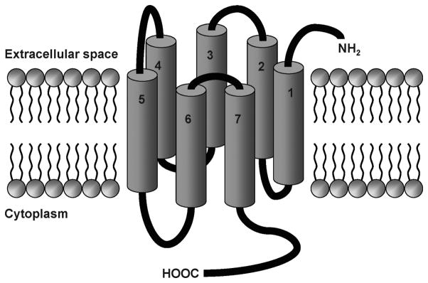
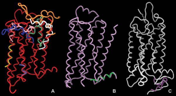
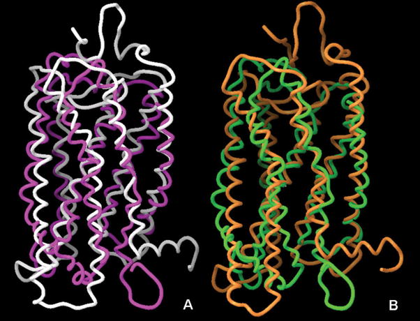
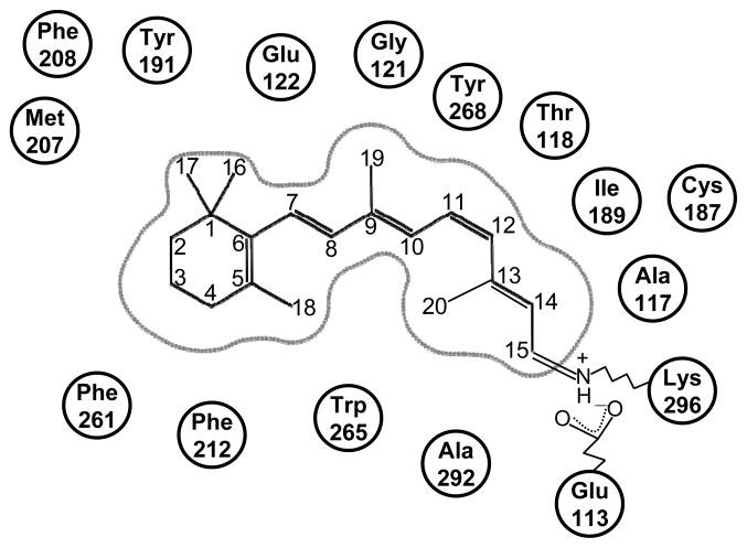
G protein-coupled receptors (GPCRs) are a large superfamily of signaling proteins expressed on the plasma membrane. They are involved in a wide range of physiological processes and, therefore, are exploited as drug targets in a multitude of therapeutic areas. In this extent, knowledge of structural and functional properties of GPCRs may greatly facilitate rational design of modulator compounds. Solution and solid-state nuclear magnetic resonance (NMR) spectroscopy represents a powerful method to gather atomistic insights into protein structure and dynamics. In spite of the difficulties inherent the solution of the structure of membrane proteins through NMR, these methods have been successfully applied, sometimes in combination with molecular modeling, to the determination of the structure of GPCR fragments, the mapping of receptor-ligand interactions, and the study of the conformational changes associated with the activation of the receptors. In this review, we provide a summary of the NMR contributions to the study of the structure and function of GPCRs, also in light of the published crystal structures.
Figures
References
-
- Pierce KL, Premont RT, Lefkowitz RJ. Seven-transmembrane receptors. Nature Reviews Molecular Cell Biology. 2002;3:639–650. - PubMed
-
- Takeda S, Kadowaki S, Haga T, Takaesu H, Mitaku S. Identification of G protein-coupled receptor genes from the human genome sequence. Febs Letters. 2002;520:97–101. - PubMed
-
- Yeagle PL, Albert AD. G-protein coupled receptor structure. Biochimica et Biophysica Acta-Biomembranes. 2007;1768:808–824. - PubMed
-
- Li J, Edwards PC, Burghammer M, Villa C, Schertler GF. Structure of bovine rhodopsin in a trigonal crystal form. J Mol Biol. 2004;343:1409–1438. - PubMed
-
- Okada T, Sugihara M, Bondar AN, Elstner M, Entel P, Buss V. The retinal conformation and its environment in rhodopsin in light of a new 2.2 Å crystal structure. J Mol Biol. 2004;342:571–583. - PubMed
Publication types
MeSH terms
Substances
Grants and funding
LinkOut - more resources
Full Text Sources
