

Role of tumor-associated lymphatic endothelial cells in metastasis: a study of epithelial ovarian tumor in vitro
- PMID: 20028387
- PMCID: PMC11158915
- DOI: 10.1111/j.1349-7006.2009.01436.x
Role of tumor-associated lymphatic endothelial cells in metastasis: a study of epithelial ovarian tumor in vitro
Abstract
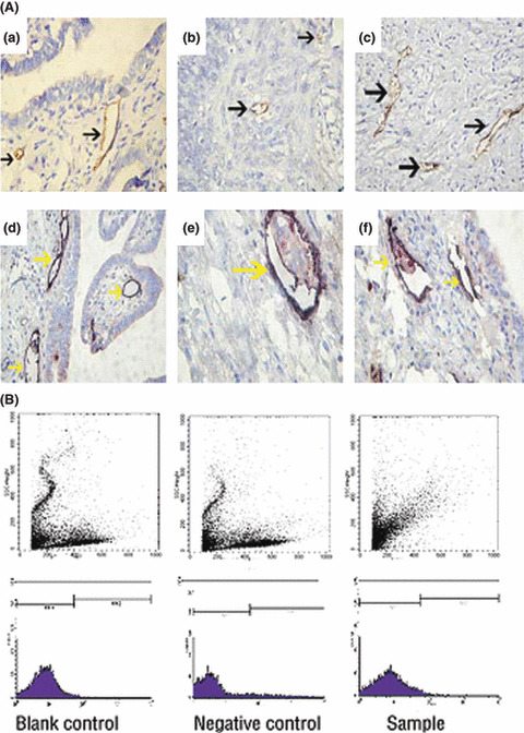
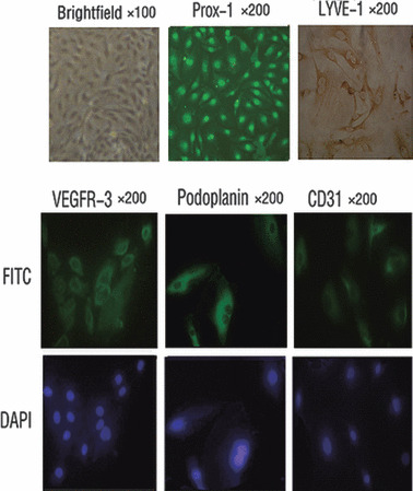
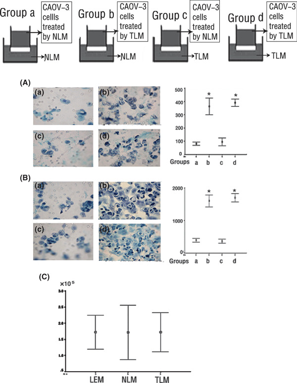
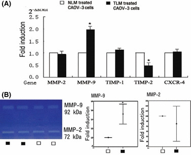
Tumor-associated lymphatic endothelial cells (TLEC) could play a key role in the process of tumor metastasis. The aim of this study was to investigate the effect of TLECs that were isolated from human epithelial ovarian tumor (EOT) on ovarian cancer cell line CAOV-3 in vitro. First, TLECs in EOT were detected by immunochemistry and flow cytometry, then marked by lymphatic endothelial cell (LEC) marker LYVE-1, isolated by magnetic beads, and cultured in vitro. The cells were identified by immunostaining of LEC markers LYVE-1, Prox-1, Podoplanin, VEGFR-3, and pan-endothelial cell marker CD31. TLECs from EOT can be detected, cultured, and identified in vitro successfully. The effects of TLECs on invasion and migration of CAOV-3 cells were investigated by 12-well Boyden chamber; the proliferation effect was studied by counting the Trypan blue exclusion cell number. Furthermore, changes in MMP-2/9 secreted by CAOV-3 cells treated with TLEC were shown using real-time PCR and zymography, and TIMP-1/2 was detected by real-time PCR. In vitro, TLECs can enhance invasion and migration of CAOV-3 cells, but have no significant effect on proliferation. It was clear that the expression of MMP-9 increased and TIMP-2 decreased in CAOV-3 cells treated by TLECs, and the increasing of MMP-9 was confirmed by zymography. TLECs from EOT can enhance migration and invasion of human ovarian carcinoma cell line in vitro, and the possible mechanism was through activation of MMP-9/TIMP-2.
Figures
Similar articles
-
Tumor-associated lymphatic endothelial cell promotes invasion of cervical cancer cells.APMIS. 2013 Dec;121(12):1162-8. doi: 10.1111/apm.12068. Epub 2013 Apr 9. APMIS. 2013. PMID: 23566114
-
Role of CXCL12 in metastasis of human ovarian cancer.Chin Med J (Engl). 2007 Jul 20;120(14):1251-5. Chin Med J (Engl). 2007. PMID: 17697577
-
[Correlation of expression of vascular endothelial growth factor and matrix matalloproteinase-2 to invasion of ovarian tumor cells in vitro].Ai Zheng. 2002 Mar;21(3):263-6. Ai Zheng. 2002. PMID: 12451991 Chinese.
-
Expression of matrix metalloproteinases (MMP-2, MMP-9, MT1-MMP) and their inhibitors (TIMP-1, TIMP-2) in common epithelial tumors of the ovary.Int J Oncol. 2000 Oct;17(4):673-81. Int J Oncol. 2000. PMID: 10995877
-
Pleuromutilin Inhibits Proliferation and Migration of A2780 and Caov-3 Ovarian Carcinoma Cells and Growth of Mouse A2780 Tumor Xenografts by Down-Regulation of pFAK2.Med Sci Monit. 2020 Feb 11;26:e920407. doi: 10.12659/MSM.920407. Med Sci Monit. 2020. Retraction in: Med Sci Monit. 2021 Jan 21;27:e930778. doi: 10.12659/MSM.930778. PMID: 32041931 Free PMC article. Retracted.
Cited by
-
Podoplanin (PDPN) affects the invasiveness of thyroid carcinoma cells by inducing ezrin, radixin and moesin (E/R/M) phosphorylation in association with matrix metalloproteinases.BMC Cancer. 2019 Jan 17;19(1):85. doi: 10.1186/s12885-018-5239-z. BMC Cancer. 2019. PMID: 30654768 Free PMC article.
-
First evaluation of [68Ga]Ga-NOTA-(TMVP1)2 for imaging VEGFR-3 in ovarian cancer patients.Am J Nucl Med Mol Imaging. 2025 Apr 15;15(2):65-73. doi: 10.62347/FYYB3942. eCollection 2025. Am J Nucl Med Mol Imaging. 2025. PMID: 40401112 Free PMC article.
-
Identification of tumour-reactive lymphatic endothelial cells capable of inducing progression of gastric cancer.Br J Cancer. 2015 Sep 29;113(7):1046-54. doi: 10.1038/bjc.2015.282. Epub 2015 Sep 10. Br J Cancer. 2015. PMID: 26355233 Free PMC article.
References
-
- Wilting J, Papoutsi M, Christ B et al. The transcription factor Prox1 is a marker for lymphatic endothelial cells in normal and diseased human tissues. FASEB J 2002; 16: 1271–3. - PubMed
-
- Dumoff KL, Chu C, Xu X et al. D2‐40 immunoreactivity correlates with lymphatic invasion and nodal metastasis in early‐stage squamous cell carcinoma of the uterine cervix. Mod Pathol 2005; 18: 97–104. - PubMed
-
- He Y, Karpanen T, Alitalo K. Role of lymphangiogenic factors in tumor metastasis. Biochim Biophys Acta 2004; 1654: 3–12. - PubMed
Publication types
MeSH terms
Substances
LinkOut - more resources
Full Text Sources
Medical
Research Materials
Miscellaneous
