. 2009 Dec 22:10:95.
doi: 10.1186/1471-2121-10-95.

Identification of MarvelD3 as a tight junction-associated transmembrane protein of the occludin family

Affiliations

Identification of MarvelD3 as a tight junction-associated transmembrane protein of the occludin family

Emily Steed et al. BMC Cell Biol. .

Abstract

Background: Tight junctions are an intercellular adhesion complex of epithelial and endothelial cells, and form a paracellular barrier that restricts the diffusion of solutes on the basis of size and charge. Tight junctions are formed by multiprotein complexes containing cytosolic and transmembrane proteins. How these components work together to form functional tight junctions is still not well understood and will require a complete understanding of the molecular composition of the junction.

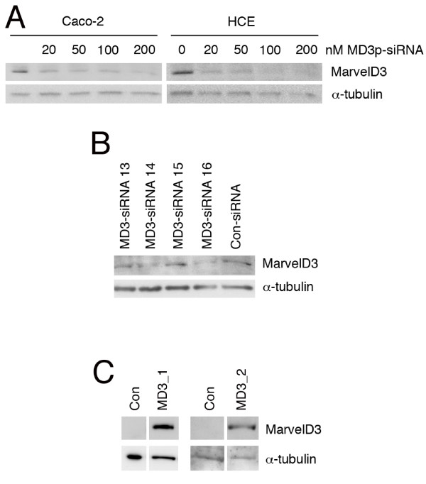
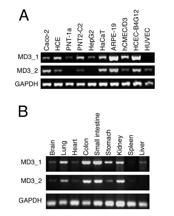
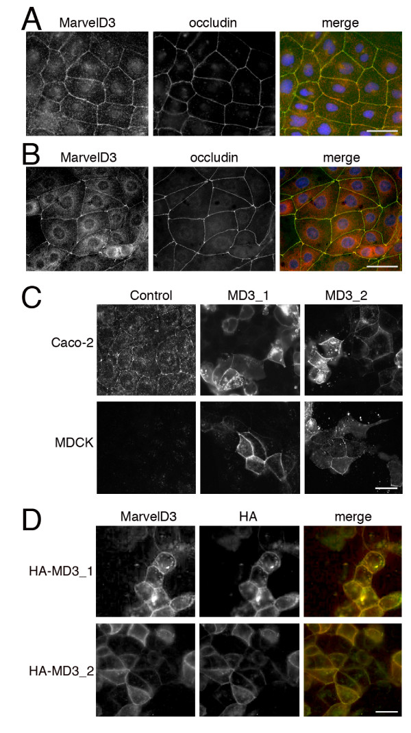
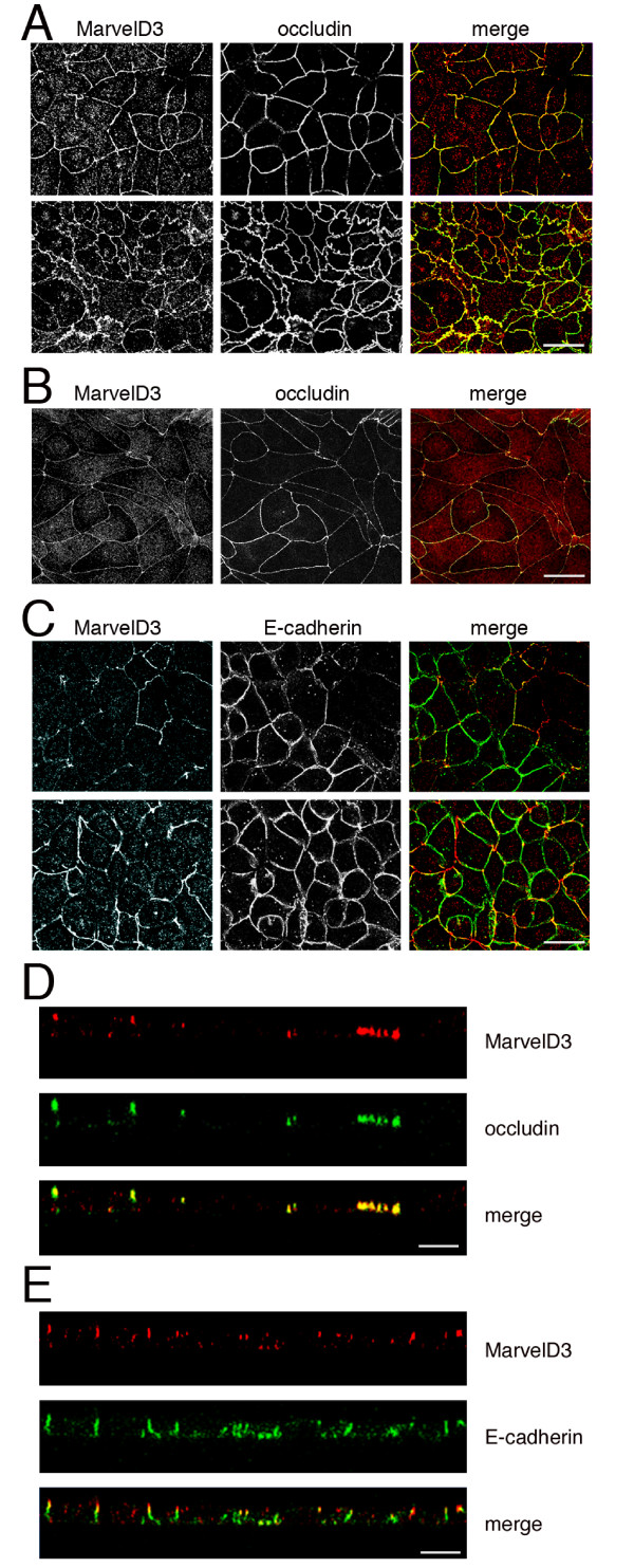
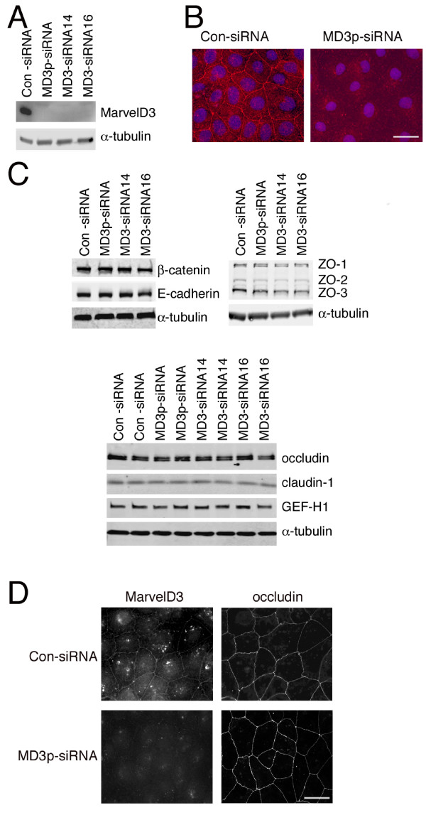
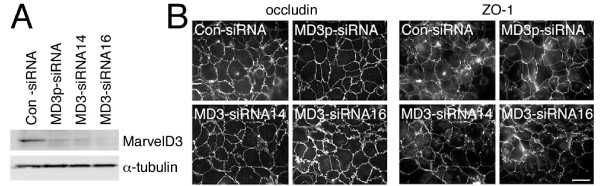
Results: Here we identify a new transmembrane component of tight junctions: MarvelD3, a four-span transmembrane protein. Its predicted transmembrane helices form a Marvel (MAL and related proteins for vesicle traffic and membrane link) domain, a structural motif originally discovered in proteins involved in membrane apposition and fusion events, such as the tight junction proteins occludin and tricellulin. In mammals, MarvelD3 is expressed as two alternatively spliced isoforms. Both isoforms exhibit a broad tissue distribution and are expressed by different types of epithelial as well as endothelial cells. MarvelD3 co-localises with occludin at tight junctions in intestinal and corneal epithelial cells. RNA interference experiments in Caco-2 cells indicate that normal MarvelD3 expression is not required for the formation of functional tight junctions but depletion results in monolayers with increased transepithelial electrical resistance.

Conclusions: Our data indicate that MarvelD3 is a third member of the tight junction-associated occludin family of transmembrane proteins. Similar to occludin, normal expression of MarvelD3 is not essential for the formation of functional tight junctions. However, MarvelD3 functions as a determinant of epithelial paracellular permeability properties.


Figures

Figure 1
Figure 1
Analysis of vertebrate MarvelD3 sequences. (A) Human MarvelD3 isoforms; (B) human, mouse and dog isoform 1 and chicken variant B; and (C) human, mouse and dog isoform 2 and chicken, Xenopus and zebrafish variant A were aligned with ClustalW http://www.ebi.ac.uk/Tools/clustalw2/ using default settings. In panel A, the cytosolic domains are highlighted in blue, the transmembrane domains in orange, and the extracellular domains in green. The splice junction between the N-terminal domain shared by both isoforms and the alternative domains is indicated by two arrows. In panels B and C, the amino acid residues conserved in mammalian MarvelD3 sequences are highlighted in yellow. Conservation is labelled according to ClustalW definitions: identical residues (*); conserved substitutions (:); semi-conserved substitutions (.).
Figure 2
Figure 2
Expression of MarvelD3 in epithelial cell lines. (A) Endogenous levels of MarvelD3 proteins in Caco-2 and HCE cells. The two cell lines were transfected with the indicated concentrations of siRNAs, using a pool of the four MarvelD3-directed siRNAs. After cell lysis, expression levels of MarvelD3 and α-tubulin were analysed by immunoblotting. (B) Identification of functional siRNAs. Caco-2 cells were transfected with individual siRNAs targeting MarvelD3 or control siRNAs. Depletion of MarvelD3 was then analysed by immunoblotting. (C) Exogenous expression of MarvelD3 isoforms. Caco-2 cells were transfected with cDNAs encoding either isoform 1 or isoform 2 of MarvelD3. Expression was then analyzed by immunoblotting with anti-MarvelD3 antibody. MD3_1 and MD3_2 constructs were run on separate SDS-PAGE gels and are shown alongside control transfections run on the same gels. Note, endogenous levels of MarvelD3 are only detected at longer exposures than those used to detect transfected proteins.
Figure 3
Figure 3
Expression of MarvelD3 isoforms in different epithelial and endothelial cells. Reverse transcription PCR was used to analyse the expression of MarvelD3 isoforms in cultured human epithelial and endothelial cells (A) and adult mouse tissues (B). Primers were used to specifically amplify MarvelD3 isoforms or, as a control, GAPDH. In panel A, cell lines and primary cultures derived from the following cell types were used: Caco-2, colon adenocarcinoma cells; HCE, immortalised corneal epithelial cells; PNT-1a and PNT2-C2, immortalised prostate epithelial cells; HepG2, hepatocellular carcinoma cells; HaCaT, spontaneously immortalised skin keratinocytes; ARPE-19, spontaneously immortalised retinal pigment epithelial cells; hCMEC/D3, immortalised brain endothelial cells; HCEC-B4G12, immortalised corneal endothelial cells; HUVEC, umbilical vein endothelial cells. Note, mRNAs for both isoforms are widely expressed by epithelial and endothelial cells.
Figure 4
Figure 4
Localisation of MarvelD3 by epifluorescence microscopy. (A, B) Immunostaining of Caco-2 (A) and HCE (B) cells with anti-MarvelD3 and anti-occludin antibodies. Cells were cultured on glass coverslips and fixed with methanol. (C) Localisation of MarvelD3 constructs in Caco-2 and MDCK cells. Caco-2 and MDCK cells were transfected with full length constructs of MarvelD3 isoform 1 (MD3_1) and isoform 2 (MD3_2). The samples were then fixed with methanol and labelled with the anti-MarvelD3 antibody. Note, both isoforms are equally distributed and localise to cell junctions. (D) Expression of HA-tagged MarvelD3. Caco-2 cells were transfected with HA-tagged variants of the MarvelD3 isoforms and then double labelled with anti-HA and anti-MarvelD3 antibodies. Bars, 10 μm.
Figure 5
Figure 5
Localisation of MarvelD3 by confocal microscopy. Caco-2 cells grown on filters (A, C, D, E) and HCE (B) cells grown on coverslips were fixed and processed for immunofluorescence with the indicated antibodies. The samples were then analyzed by confocal microscopy. Panels A, B, and C are xy sections, and D and E are reconstitutions of serial z line scans. In panels A and C, two sections from different samples are shown that were both taken at the interface between the tight and adherens junctions to facilitate comparison between the two different labels in each specimen. Note, occludin and MarvelD3 tightly follow each other in and out of the focal plane. Bars, 10 μm.
Figure 6
Figure 6
Depletion of MarvelD3 in Caco-2 cells. Caco-2 cells were transfected with the indicated siRNAs and then processed for immunoblotting (A and C) or immunofluorescence (B and D). (A, C) Cells were immunoblotted with antibodies against MarvelD3 and α-tubulin (A) or against a selection of tight and adherens junction proteins as indicated. Note, the expression levels of none of the junctional proteins apart from MarvelD3 were affected by depletion of the latter protein independent of whether filter- or glass-grown cells were analysed. Bar, 10 μm. (C) The lower panel in C shows duplicate cell extracts for each type of siRNA transfection. (B) Immunofluorescence staining of cells labelled with anti-MarvelD3 antibodies and Hoechst dye to stain DNA. Note, reduced levels of MarvelD3 were seen following transfection with MarvelD3 siRNAs. (D) siRNA transfected cells were labelled with anti-MavelD3 and anti-occludin antibodies. Bar, 10 μm. Note, knockdown of MarvelD3 did not appear to affect occludin distribution.
Figure 7
Figure 7
Depletion of MarvelD3 and tight junction assembly. (A) Control and siRNA-transfected Caco-2 cells were plated on filters one day after transfection. The cells were lysed three days later and expression of MarvelD3 and α-tubulin was determined by immunoblotting. (B) Cells treated as those in panel A were fixed and processed for immunofluorescence at the end of the incubation period. Shown are epifluorescence images of samples labelled for the tight junction markers occludin and ZO-1. Bar, 10 μm. Note, depletion of MarvelD3 did not affect monolayer integrity and appearance.
Figure 8
Figure 8
Depletion of MarvelD3 and epithelial barrier properties. Cells were cultured as in fig. 5 either using the Ca2+ Switch (A, B) or Direct plating (C, D) protocol. TER and fluorescent dextran permeability using 4 kD and 70 kD dextran were measured as indicated. The amount of dextran diffused to the basolateral side of the monolayer was normalised against the average value obtained from control cells. Shown are averages ± 1SD of quadruplicate samples of a typical experiment. The indicated p values were obtained with a t-test comparing knockdown with control values; in panel A, the p values refer to the final TER values. Note, MarvelD3 knockdown had no significant effect on diffusion of either dextran tracer across monolayers in either the Ca2+ switch experiment (B) or those plated directly into complete culture medium (D). The apparent decreases in the mean values in panel B obtained for single siRNA transfections were neither statistically significant nor did they reflect a trend observed in other experiments. However, final TER values were elevated in both culture conditions in all performed experiments.

Similar articles

Cited by

References

    1. Cereijido M, Contreras RG, Shoshani L. Cell adhesion, polarity, and epithelia in the dawn of metazoans. Physical rev. 2004;84(4):1229–1262. doi: 10.1152/physrev.00001.2004. - DOI - PubMed
    1. Tsukita S, Furuse M, Itoh M. Multifunctional strands in tight junctions. Nat Rev Mol Cell Biol. 2001;2:286–293. doi: 10.1038/35067088. - DOI - PubMed
    1. Aijaz S, Balda MS, Matter K. Tight Junctions: Molecular Architecture and Function. Int Rev Cytol. 2006;248:261–298. doi: 10.1016/S0074-7696(06)48005-0. - DOI - PubMed
    1. Chiba H, Osanai M, Murata M, Kojima T, Sawada N. Transmembrane proteins of tight junctions. Biochim Biophys Acta. 2008;1778(3):588–600. doi: 10.1016/j.bbamem.2007.08.017. - DOI - PubMed
    1. Bazzoni G. The JAM family of junctional adhesion molecules. Curr Opin Cell Biol. 2003;15(5):525–530. doi: 10.1016/S0955-0674(03)00104-2. - DOI - PubMed

Publication types