

Expression of classic cadherins and delta-protocadherins in the developing ferret retina
- PMID: 20028529
- PMCID: PMC2811116
- DOI: 10.1186/1471-2202-10-153
Expression of classic cadherins and delta-protocadherins in the developing ferret retina
Abstract
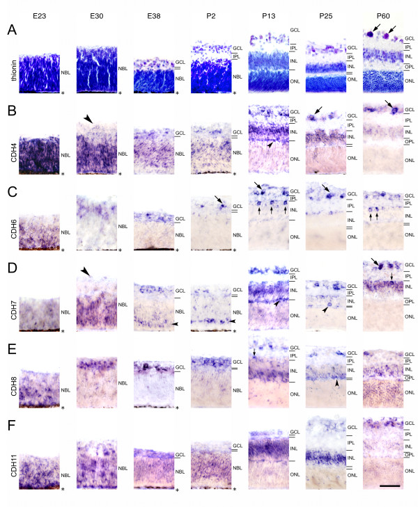
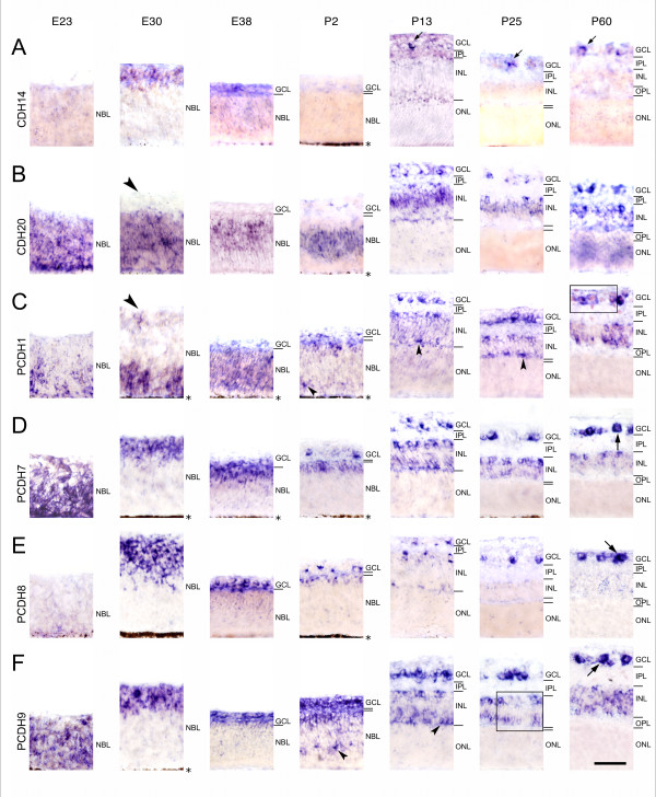
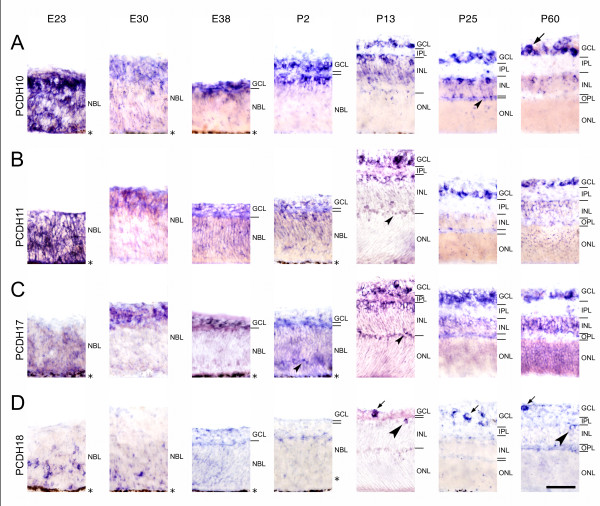
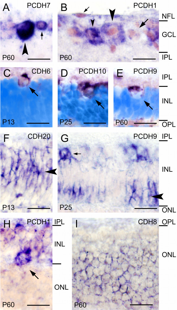
Background: Cadherins are a superfamily of calcium-dependent adhesion molecules that play multiple roles in morphogenesis, including proliferation, migration, differentiation and cell-cell recognition. The subgroups of classic cadherins and delta-protocadherins are involved in processes of neural development, such as neurite outgrowth, pathfinding, target recognition, synaptogenesis as well as synaptic plasticity. We mapped the expression of 7 classic cadherins (CDH4, CDH6, CDH7, CDH8, CDH11, CDH14, CDH20) and 8 delta-protocadherins (PCDH1, PCDH7, PCDH8, PCDH9, PCDH10, PCDH11, PCDH17, PCDH18) at representative stages of retinal development and in the mature retina of the ferret by in situ hybridization.
Results: All cadherins investigated by us are expressed differentially by restricted populations of retinal cells during specific periods of the ferret retinogenesis. For example, during embryonic development, some cadherins are exclusively expressed in the outer, proliferative zone of the neuroblast layer, whereas other cadherins mark the prospective ganglion cell layer or cells in the prospective inner nuclear layer. These expression patterns anticipate histogenetic changes that become visible in Nissl or nuclear stainings at later stages. In parallel to the ongoing development of retinal circuits, cadherin expression becomes restricted to specific subpopulations of retinal cell types, especially of ganglion cells, which express most of the investigated cadherins until adulthood. A comparison to previous results in chicken and mouse reveals overall conserved expression patterns of some cadherins but also species differences.
Conclusions: The spatiotemporally restricted expression patterns of 7 classic cadherins and 8 delta-protocadherins indicate that cadherins provide a combinatorial adhesive code that specifies developing retinal cell populations and intraretinal as well as retinofugal neural circuits in the developing ferret retina.
Figures
Similar articles
-
Cadherin expression in the somatosensory cortex: evidence for a combinatorial molecular code at the single-cell level.Neuroscience. 2011 Feb 23;175:37-48. doi: 10.1016/j.neuroscience.2010.11.056. Epub 2010 Dec 1. Neuroscience. 2011. PMID: 21129452
-
Cadherin expression delineates the divisions of the postnatal and adult mouse amygdala.J Comp Neurol. 2012 Dec 1;520(17):3982-4012. doi: 10.1002/cne.23140. J Comp Neurol. 2012. PMID: 22592879
-
Expression of delta-protocadherins in the spinal cord of the chicken embryo.J Comp Neurol. 2012 May 1;520(7):1509-31. doi: 10.1002/cne.22808. J Comp Neurol. 2012. PMID: 22102158
-
Non-clustered protocadherin.Cell Adh Migr. 2011 Mar-Apr;5(2):97-105. doi: 10.4161/cam.5.2.14374. Epub 2011 Mar 1. Cell Adh Migr. 2011. PMID: 21173574 Free PMC article. Review.
-
Cadherins in cerebellar development: translation of embryonic patterning into mature functional compartmentalization.Cerebellum. 2011 Sep;10(3):393-408. doi: 10.1007/s12311-010-0207-4. Cerebellum. 2011. PMID: 20820976 Review.
Cited by
-
Physiological Functions of Thiol Peroxidases (Gpx1 and Prdx2) during Xenopus laevis Embryonic Development.Antioxidants (Basel). 2021 Oct 17;10(10):1636. doi: 10.3390/antiox10101636. Antioxidants (Basel). 2021. PMID: 34679770 Free PMC article.
-
Low Expression of Protocadherin-8 Promotes the Progression of Ovarian Cancer.Int J Gynecol Cancer. 2018 Feb;28(2):346-354. doi: 10.1097/IGC.0000000000001169. Int J Gynecol Cancer. 2018. PMID: 29324532 Free PMC article.
-
Analysis of gene expression during mineralization of cultured human periodontal ligament cells.J Periodontal Implant Sci. 2011 Feb;41(1):30-43. doi: 10.5051/jpis.2011.41.1.30. Epub 2011 Feb 28. J Periodontal Implant Sci. 2011. PMID: 21394295 Free PMC article.
-
Loss of flrt2 gene leads to microphthalmia in zebrafish.Biol Open. 2023 Jun 15;12(6):bio059784. doi: 10.1242/bio.059784. Epub 2023 Jun 15. Biol Open. 2023. PMID: 37259881 Free PMC article.
-
Host-virus interactions during infection with a wild-type ILTV strain or a glycoprotein G deletion mutant ILTV vaccine strain in an ex vivo system.Microbiol Spectr. 2025 Feb 4;13(2):e0118324. doi: 10.1128/spectrum.01183-24. Epub 2025 Jan 13. Microbiol Spectr. 2025. PMID: 39804092 Free PMC article.
References
Publication types
MeSH terms
Substances
LinkOut - more resources
Full Text Sources
Miscellaneous
