

Targeting of bone-derived insulin-like growth factor-II by a human neutralizing antibody suppresses the growth of prostate cancer cells in a human bone environment
- PMID: 20028742
- PMCID: PMC2802676
- DOI: 10.1158/1078-0432.CCR-09-0982
Targeting of bone-derived insulin-like growth factor-II by a human neutralizing antibody suppresses the growth of prostate cancer cells in a human bone environment
Abstract
Purpose: Advanced prostate cancer frequently involves the bone, where the insulin-like growth factor (IGF)-II is abundant. However, the importance of IGF-II in bone metastasis from prostate cancer is uncertain. The present study was aimed at examining the therapeutic importance of targeting IGF-II in bone metastases from prostate cancer.
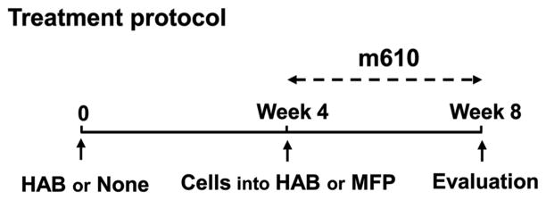
Experimental design: We investigated whether inhibiting IGF-II using a human neutralizing antibody (m610) suppresses the growth of prostate cancer cells in a human bone environment. Human MDA PCa 2b prostate cancer cells were inoculated into human adult bone implanted into mammary fat pad of nonobese diabetic/severe combined immunodeficient mice or inoculated into mammary fat pad of the mice without human bone implantation. The mice were treated with m610 or a control antibody (m102.4) once weekly for 4 weeks immediately after inoculation with MDA PCa 2b cells.
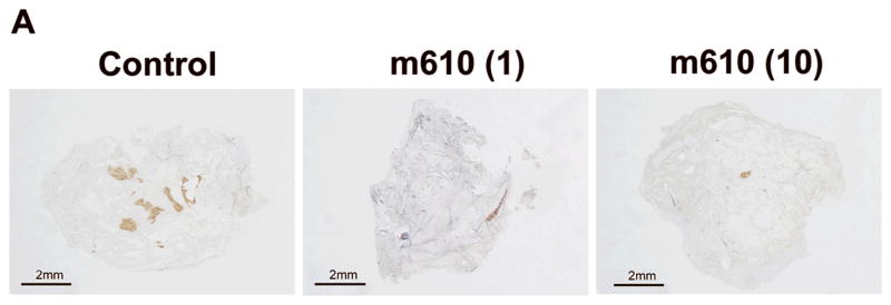
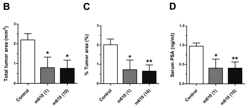
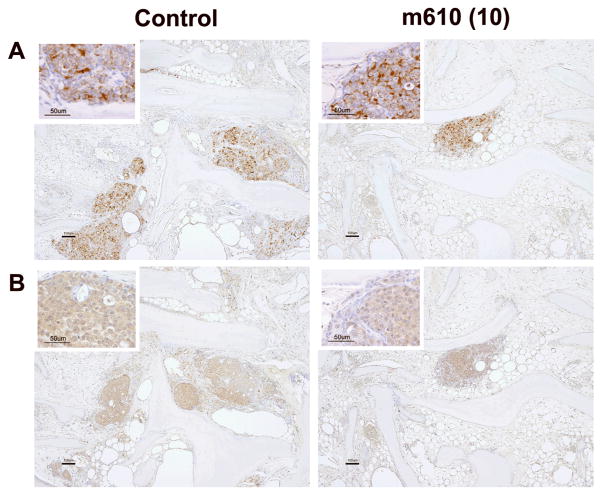
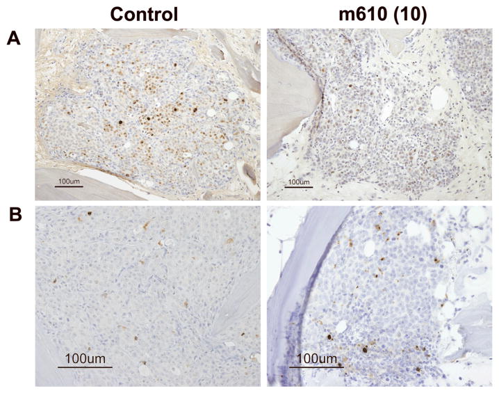
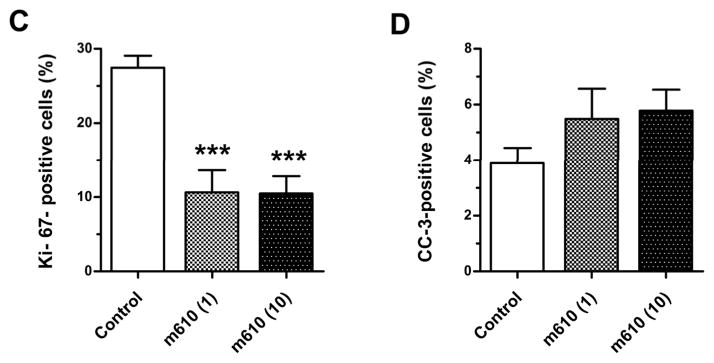
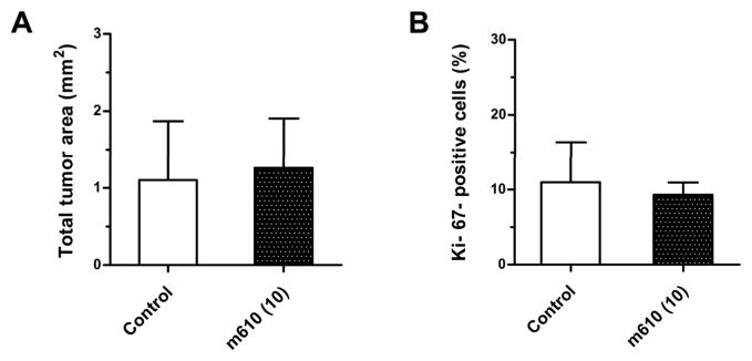
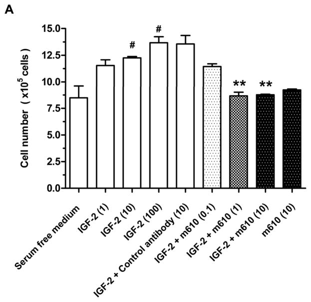
Results: Histomorphologic examination indicated that m610 treatment significantly decreased the MDA PCa 2b tumor area in the human bone compared with the control. Ki-67 immunostaining revealed that the percentage of proliferating cancer cells in the m610-treated bone tumor sections was significantly lower than that in the control. m610 had no effect on MDA PCa 2b tumor growth in the absence of implanted human bone. m610 prevented the in vitro IGF-II-induced proliferation of MDA PCa 2b cells.
Conclusions: Our results indicate that IGF-II plays an important role in the prostate cancer cell growth in human bone, suggesting that targeting it by neutralizing antibodies offers a new therapeutic strategy for bone metastasis from prostate cancer.
Figures
Similar articles
-
Growth inhibition of human prostate cancer cells in human adult bone implanted into nonobese diabetic/severe combined immunodeficient mice by a ligand-specific antibody to human insulin-like growth factors.Cancer Res. 2004 Sep 1;64(17):6252-8. doi: 10.1158/0008-5472.CAN-04-0919. Cancer Res. 2004. PMID: 15342412
-
Prostate cancer cells-osteoblast interaction shifts expression of growth/survival-related genes in prostate cancer and reduces expression of osteoprotegerin in osteoblasts.Clin Cancer Res. 2003 Jul;9(7):2587-97. Clin Cancer Res. 2003. PMID: 12855635
-
Novel human monoclonal antibodies to insulin-like growth factor (IGF)-II that potently inhibit the IGF receptor type I signal transduction function.Mol Cancer Ther. 2006 Jan;5(1):114-20. doi: 10.1158/1535-7163.MCT-05-0252. Mol Cancer Ther. 2006. PMID: 16432169
-
Insulin-Like Growth Factor (IGF) family and prostate cancer.Crit Rev Oncol Hematol. 2006 May;58(2):124-45. doi: 10.1016/j.critrevonc.2005.10.003. Epub 2006 Jan 18. Crit Rev Oncol Hematol. 2006. PMID: 16387509 Review.
-
Parathyroid hormone-related protein and bone metastases.Cancer. 1997 Oct 15;80(8 Suppl):1572-80. doi: 10.1002/(sici)1097-0142(19971015)80:8+<1572::aid-cncr7>3.3.co;2-d. Cancer. 1997. PMID: 9362424 Review.
Cited by
-
The endoplasmic reticulum protein folding factory and its chaperones: new targets for drug discovery?Br J Pharmacol. 2011 Jan;162(2):328-45. doi: 10.1111/j.1476-5381.2010.01064.x. Br J Pharmacol. 2011. PMID: 20942857 Free PMC article. Review.
-
Growth factors involve in cellular proliferation, differentiation and migration during prostate cancer metastasis.Int J Cell Biol Physiol. 2019;2(1-2):1-13. Epub 2019 Oct 7. Int J Cell Biol Physiol. 2019. PMID: 32259163 Free PMC article.
-
Ligand-independent activation of MET through IGF-1/IGF-1R signaling.Int J Cancer. 2013 Oct 1;133(7):1536-46. doi: 10.1002/ijc.28169. Epub 2013 Apr 17. Int J Cancer. 2013. PMID: 23526299 Free PMC article.
-
Collagenous and non-collagenous biochemical markers of bone metastases from prostate cancer.Hippokratia. 2010 Jul;14(3):164-9. Hippokratia. 2010. PMID: 20981164 Free PMC article.
-
A new bispecific antibody targeting non-overlapping epitopes on IGF2: design, in vitro characterization and pharmacokinetics in macaques.Exp Mol Pathol. 2014 Dec;97(3):359-67. doi: 10.1016/j.yexmp.2014.09.007. Epub 2014 Sep 16. Exp Mol Pathol. 2014. PMID: 25220345 Free PMC article.
References
-
- Abrams HL, Spiro R, Goldstein N. Metastasis in carcinoma: analysis of 1000 autopsied cases. Cancer (Phila) 1950;3:74–85. - PubMed
-
- Bubendorf L, Schopfer A, Wagner U, et al. Metastatic patterns of prostate cancer: an autopsy study of 1,589 patients. Hum Pathol. 2000;31:578–83. - PubMed
-
- Soloway MS. The importance of prognostic factors in advanced prostate cancer. Cancer. 1990;66:1017–21. - PubMed
-
- Yoneda T. Cellular and molecular mechanisms of breast and prostate cancer metastasis to bone. Eur J Cancer. 1998;34:240–5. - PubMed
-
- Yonou H, Yokose T, Kamijo T, et al. Establishment of a novel species- and tissue-specific metastasis model of human prostate cancer in humanized non-obese diabetic/severe combined immunodeficient mice engrafted with human adult lung and bone. Cancer Res. 2001;61:2177–82. - PubMed
Publication types
MeSH terms
Substances
Grants and funding
LinkOut - more resources
Full Text Sources
Other Literature Sources
Medical
