. 2010 Jan 1;16(1):45-55.
doi: 10.1158/1078-0432.CCR-09-1630. Epub 2009 Dec 22.

Aldehyde dehydrogenase 1-positive cancer stem cells mediate metastasis and poor clinical outcome in inflammatory breast cancer

Affiliations

Aldehyde dehydrogenase 1-positive cancer stem cells mediate metastasis and poor clinical outcome in inflammatory breast cancer

Emmanuelle Charafe-Jauffret et al. Clin Cancer Res. .

Abstract

Purpose: To examine the role of cancer stem cells (CSC) in mediating metastasis in inflammatory breast cancer (IBC) and the association of these cells with patient outcome in this aggressive type of breast cancer.

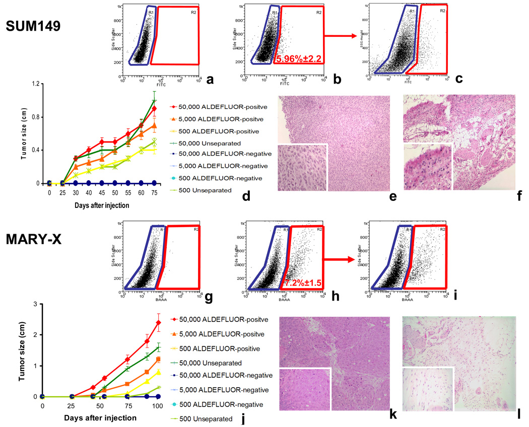
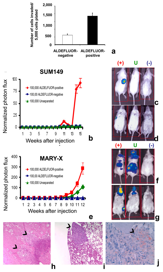
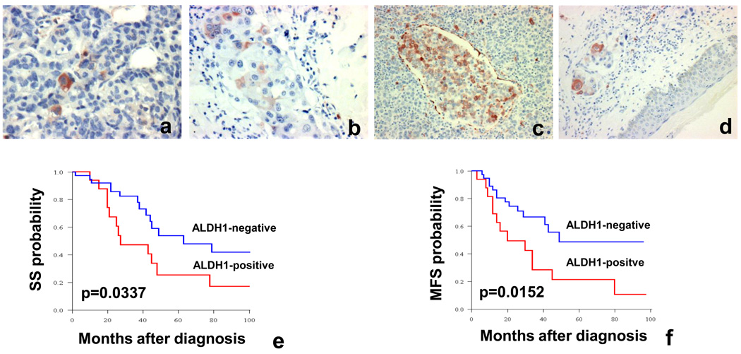
Experimental design: CSCs were isolated from SUM149 and MARY-X, an IBC cell line and primary xenograft, by virtue of increased aldehyde dehydrogenase (ALDH) activity as assessed by the ALDEFLUOR assay. Invasion and metastasis of CSC populations were assessed by in vitro and mouse xenograft assays. Expression of ALDH1 was determined on a retrospective series of 109 IBC patients and this was correlated with histoclinical data. All statistical tests were two sided. Log-rank tests using Kaplan-Meier analysis were used to determine the correlation of ALDH1 expression with development of metastasis and patient outcome.

Results: Both in vitro and xenograft assays showed that invasion and metastasis in IBC are mediated by a cellular component that displays ALDH activity. Furthermore, expression of ALDH1 in IBC was an independent predictive factor for early metastasis and decreased survival in this patient population.

Conclusions: These results suggest that the metastatic, aggressive behavior of IBC may be mediated by a CSC component that displays ALDH enzymatic activity. ALDH1 expression represents the first independent prognostic marker to predict metastasis and poor patient outcome in IBC. The results illustrate how stem cell research can translate into clinical practice in the IBC field.


Figures

Figure 1
Figure 1. The ALDEFLUOR-positive cell population of SUM149 and MARY-X cells displays properties of cancer stem cells
a–b, g–h. Representative flow cytometry analysis of ALDH activity in SUM149 (a–b) and MARY-X (g–h) inflammatory breast carcinoma cells. Cells were incubated with ALDEFLUOR substrate (BAAA) and the specific inhibitor of ALDH, DEAB, was used to establish the baseline fluorescence of these cells (R1) and to define the ALDEFLUOR-positive region (R2) (a, g). Incubation of cells with ALDEFLUOR substrate in the absence of DEAB induces a shift in BAAA fluorescence defining the ALDEFLUOR-positive population which represents 5.96±2.2% in SUM149 and 7.2±1.5% in MARY-X of the total population (b, h). All of the ALDEFLUOR analyses on human breast tumor cells were first gated on PI negative cells (viable cells) which represented 99.98± 0.0282% (Mean ± SDEV, n=7) of the total population. c–f, i–l. In the two models utilized, only the ALDEFLUOR-positive population was tumorigenic. c, i. The ALDEFLUOR-positive population was capable of regenerating the phenotypic heterogeneity of the initial tumor after passage in NOD/SCID mice. d, j. For SUM149 and MARY-X, varying numbers of ALDEFLUOR-positive and ALDEFLUOR-negative cells were injected and tumor growth was measured over a 75 days interval for SUM149 and a 100 days interval for MARY-X. No tumor was detected when 50,000 ALDEFLUOR-negative cells were injected, whereas ALDEFLUOR-positive cells produced tumors which grew at a rate that directly correlated with the number of cells injected. Similar results were observed for SUM149 and MARY-X. e–f, k–l. H&E staining demonstrating presence of tumors at the ALDEFLUOR-positive injection site (e:SUM149; k: MARY-X) and an absence of tumor at the ALDEFLUOR-negative injection site (f:SUM149; l: MARY-X).
Figure 2
Figure 2. The ALDEFLUOR-positive cell population of SUM149 and MARY-X cells mediates invasion and metastasis
a. The ALDEFLUOR-positive population from SUM149 is associated with greater invasion potential: SUM149 ALDEFLUOR-positive cells demonstrated a three-fold increase in Matrigel invasion assay compared to SUM149 ALDEFLUOR-negative cells (p<0.05). b–i Utilizing two different models (SUM149 and MARY-X) we demonstrated that only the ALDEFLUOR-positive population displayed metastatic potential. b,e Quantification of the normalized photon flux measured at weekly intervals following intracardiac inoculations 100,000 luciferase infected cells from each group (ALDEFLUOR-positive, ALDEFLUOR-negative, unseparated) and for both models, SUM149 (b) and MARY-X (e). c–d, e–f Detection of metastasis utilizing bioluminescence imaging software. Mice injected with 100,000 SUM149 or MARY-X ALDEFLUOR-positive cells but not with ALDEFLUOR-negative cells develop systemic metastasis. h–i Histologic confirmation, on H&E sections, of metastasis in bone and lung resulting from injection of SUM149 ALDEFLUOR-positive cells (arrows). Similarly, the presence of metastases was confirmed by histologic inspection in mice inoculated with MARY-X cells. j Mary-X metastasis (spine bone) formed from intracardiac injection of ALDEFLUOR-positive cells contained cells expressing (arrow) or not ALDH1 and recapitulates heterogeneity of the initial tumor.
Figure 3
Figure 3. ALDH1 expression in IBC patient tumors is associated with the development of metastasis and with decreased survival
a–d: Example of ALDH1 expression in a subset of cells in two different IBC samples. c–d: Tumor emboli in dermal lymphatics show cells expressing ALDH1. e–f: Kaplan-Meier survival curves according to ALDH1 status. ALDH1 expression is associated with decreased SS and MFS.

Similar articles

Cited by

References

    1. Hance KW, Anderson WF, Devesa SS, Young HA, Levine PH. Trends in inflammatory breast carcinoma incidence and survival: the surveillance, epidemiology, and end results program at the National Cancer Institute. J Natl Cancer Inst. 2005;97:966–975. - PMC - PubMed
    1. Ravdin PM, Cronin KA, Howlader N, et al. The decrease in breast-cancer incidence in 2003 in the United States. N Engl J Med. 2007;356:1670–1674. - PubMed
    1. Yang CH, Cristofanilli M. The role of p53 mutations as a prognostic factor and therapeutic target in inflammatory breast cancer. Future Oncol. 2006;2:247–255. - PubMed
    1. Charafe-Jauffret E, Tarpin C, Bardou VJ, et al. Immunophenotypic analysis of inflammatory breast cancers: identification of an "inflammatory signature". J Pathol. 2004;202:265–273. - PubMed
    1. Kleer CG, Zhang Y, Pan Q, et al. WISP3 and RhoC guanosine triphosphatase cooperate in the development of inflammatory breast cancer. Breast Cancer Res. 2004;6:R110–R115. - PMC - PubMed

Publication types

MeSH terms