. 2010 Jan 1;16(1):109-20.
doi: 10.1158/1078-0432.CCR-09-2151. Epub 2009 Dec 22.

Targeting the cyclin E-Cdk-2 complex represses lung cancer growth by triggering anaphase catastrophe

Affiliations

Targeting the cyclin E-Cdk-2 complex represses lung cancer growth by triggering anaphase catastrophe

Fabrizio Galimberti et al. Clin Cancer Res. .

Abstract

Purpose: Cyclin-dependent kinases (Cdk) and their associated cyclins are targets for lung cancer therapy and chemoprevention given their frequent deregulation in lung carcinogenesis. This study uncovered previously unrecognized consequences of targeting the cyclin E-Cdk-2 complex in lung cancer.

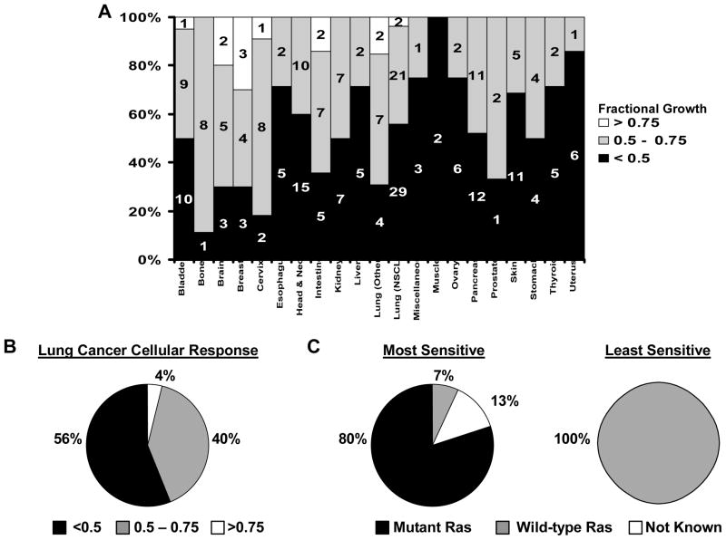
Experimental design: Cyclin E, Cdk-1, and Cdk-2 were individually targeted for repression with siRNAs in lung cancer cell lines. Cdk-2 was also pharmacologically inhibited with the reversible kinase inhibitor seliciclib. Potential reversibility of seliciclib effects was assessed in washout experiments. Findings were extended to a large panel of cancer cell lines using a robotic-based platform. Consequences of cyclin E-Cdk-2 inhibition on chromosome stability and on in vivo tumorigenicity were explored as were effects of combining seliciclib with different taxanes in lung cancer cell lines.

Results: Targeting the cyclin E-Cdk-2 complex, but not Cdk-1, resulted in marked growth inhibition through the induction of multipolar anaphases triggering apoptosis. Treatment with the Cdk-2 kinase inhibitor seliciclib reduced lung cancer formation in a murine syngeneic lung cancer model and decreased immunohistochemical detection of the proliferation markers Ki-67 and cyclin D1 in lung dysplasia spontaneously arising in a transgenic cyclin E-driven mouse model. Combining seliciclib with a taxane resulted in augmented growth inhibition and apoptosis in lung cancer cells. Pharmacogenomic analysis revealed that lung cancer cell lines with mutant ras were especially sensitive to seliciclib.

Conclusions: Induction of multipolar anaphases leading to anaphase catastrophe is a previously unrecognized mechanism engaged by targeting the cyclin E-Cdk-2 complex. This exerts substantial antineoplastic effects in the lung.


Figures

Fig. 1
Fig. 1
Individual siRNA-mediated knock-down of cyclin E species repressed growth of ED-1 and ED-2 lung cancer cell lines. A) Confirmation of cyclin E mRNA knock-down by real-time RT-PCR assays performed on RNA isolated from ED-1 (left panel) and ED-2 (right panel) cells transfected with different siRNAs targeting both human and murine cyclin E species or with RISC-free siRNA (control). B) Proliferation of ED-1 (left panel) and ED-2 (right panel) cells was inhibited by these siRNAs targeting cyclin E. Standard deviation bars are shown.
Fig. 2
Fig. 2
Cdk-2 inhibition repressed proliferation and clonal growth of ED-1 and ED-2 lung cancer cells. A) Dose-dependent and time-dependent inhibition of ED-1 (left panel) and ED-2 (right panel) cell growth by seliciclib as compared to vehicle (DMSO) control. B) Seliciclib treatment reduced clonal growth of ED-1 cells in a dose-dependent manner. C) Dose-dependent effects of pharmacological inhibition of Cdk-2 with seliciclib resulted in down-regulation of cyclin D1 protein by immunoblot analysis of ED-1 cells. RNA-polymerase II phosphorylation was inhibited by seliciclib treatment only at a dosage of 25μM (a dosage above those examined for anti-neoplastic effects). Actin expression served as a loading control. D) Seliciclib effects were partially reversed in ED-1 (left panel) and ED-2 (right panel) cells as seen in wash-out experiments at the 10μM dosage, as shown in this figure. ED-1 and ED-2 cell growth only partially recovered from seliciclib wash-out as compared to cells continuously treated with seliciclib. Standard deviation bars and p-values are displayed.
Fig. 3
Fig. 3
Cdk-2 inhibition affects chromosomal stability by inducing multipolar anaphases in murine and human lung cancer cells, but not in C-10 immortalized murine lung epithelial cells. A) A representative ED-1 cell undergoing anaphase in the presence of control (vehicle, DMSO, or control siRNA) and two indepedendent Cdk-1 siRNAs (results from one representative siRNA experiment is shown), as compared to another representative ED-1 cell undergoing multipolar anaphase in the presence of seliciclib (10μM) as well as following transfection of each of two different Cdk-2 siRNAs (results from one representative siRNA experiment is shown). ED-1 cells were fixed 24 hours after treatment and stained, as described in the Materials and Methods. Microtubules were stained red and DNA was stained blue with DAPI. Cells in anaphase were scored for mulitpolar anaphases as in the Materials and Methods. B) The left panel shows a representative induction of multipolar anaphases in ED-1 cells 24 hours after seliciclib treatments. The right panel shows induction of anaphase catastrophe in ED-1 cells 24 hours after transfection with each of two different Cdk-2 targeting siRNAs, two Cdk-1 siRNAs, and control siRNA. C–D) These panels depict the percentage of H-23, HOP-62, and H-522 human lung cancer cells as compared to C-10 immortalized murine pulmonary epithelial cells undergoing multipolar anaphases following seliciclib (15μM) treatment (0 Hours = control). Standard deviation bars and p-values are shown.
Fig. 4
Fig. 4
Seliciclib and taxanes cooperatively inhibited proliferation and clonal growth, while inducing apoptosis of ED-1 lung cancer cells. A) Seliciclib treatment combined with paclitaxel (left panel) or docetaxel (right panel) treatment cooperatively inhibited ED-1 cell growth as compared to vehicle (DMSO) controls. B) Seliciclib treatment combined with paclitaxel (left panel) or docetaxel (right panel) treatment increased apoptosis as detected by Annexin V:FITC and PI staining. C) Cooperation between seliciclib and paclitaxel (left panel) or docetaxel (right panel) treatments significantly repressed ED-1 cell clonal growth. Standard deviation bars and p-values are shown.
Fig. 5
Fig. 5
Profiling for seliciclib sensitivity revealed growth inhibitory effects in diverse cancer cell lines. A) Schematic representation of seliciclib sensitivity across 270 cancer cell lines from diverse tissues. Lung (Others) includes: 4 small cell lung cancer, 6 mesothelioma, and 1 bronchial carcinoma cell lines. Miscellaneous includes: 2 fibrosarcoma, 1 fibrous histiocytoma, and 1 small round cell sarcoma cancer cell lines. The complete set of data is presented in Supplemental Table 1. B) Pie chart representation of NSCLC cell lines sensitivity to seliciclib (15μM) treatment. C) The pie chart on the left shows ras status for the 15 NSCLC cell lines with highest growth inhibitory response to seliciclib. The pie chart on the right shows ras status for the 15 NSCLC cell lines with least growth inhibitory response to seliciclib.
Fig. 6
Fig. 6
In vivo seliciclib treatment effects. A) Seliciclib treatment caused repression of Ki-67 and cyclin D1 immunostained nuclei in representative dysplastic lung lesions of wild-type cyclin E expressing transgenic mice as compared to vehicle (DMSO) treated mice. B) Representative hematoxylin and eosin staining of lung tissues from syngeneic FVB mice injected with ED-1 lung cancer cells before in vivo treatment with seliciclib or vehicle (DMSO). C) Seliciclib treatment resulted in reduced high-grade lesions (p = 0.026) in the lungs of ED-1 tail-vein injected syngeneic FVB mice as compared to vehicle (DMSO) treatment. Each symbol represents a single mouse. The line shows the mean number of high grade lesions in each group. D) Fewer multilayer lesions (p = 0.005) occurred in the lungs of ED-1 tail-vein injected syngeneic FVB mice treated in vivo with seliciclib as compared to vehicle (DMSO) treated mice. Each symbol represents a single mouse. The line shows the mean number of multilayer lesions in each group.

Similar articles

Cited by

References

    1. Shapiro GI. Cyclin-dependent kinase pathways as targets for cancer treatment. J Clin Oncol. 2006;24:1770–83. - PubMed
    1. Koff A, Giordano A, Desai D, et al. Formation and activation of a cyclin E-cdk2 complex during the G1 phase of the human cell cycle. Science. 1992;257:1689–94. - PubMed
    1. Freemantle SJ, Liu X, Feng Q, et al. Cyclin degradation for cancer therapy and chemoprevention. J Cell Biochem. 2007;102:869–77. - PubMed
    1. Harbour JW, Luo RX, Dei Santi A, Postigo AA, Dean DC. Cdk phosphorylation triggers sequential intramolecular interactions that progressively block Rb functions as cells move through G1. Cell. 1999;98:859–69. - PubMed
    1. Ohtsubo M, Theodoras AM, Schumacher J, Roberts JM, Pagano M. Human cyclin E, a nuclear protein essential for the G1-to-S phase transition. Mol Cell Biol. 1995;15:2612–24. - PMC - PubMed

Publication types