. 2009 Dec;21(12):3950-64.
doi: 10.1105/tpc.109.069435. Epub 2009 Dec 22.

Phosphorylation of photosystem II controls functional macroscopic folding of photosynthetic membranes in Arabidopsis

Affiliations

Phosphorylation of photosystem II controls functional macroscopic folding of photosynthetic membranes in Arabidopsis

Rikard Fristedt et al. Plant Cell. 2009 Dec.

Abstract

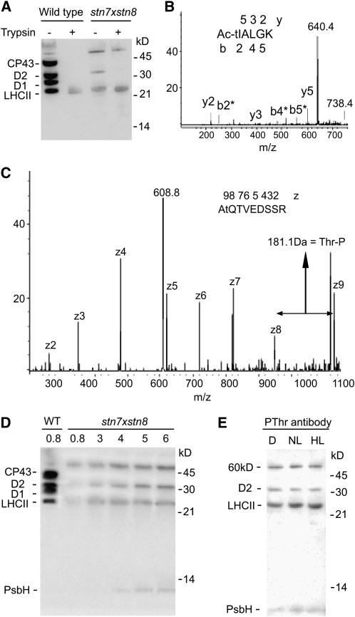
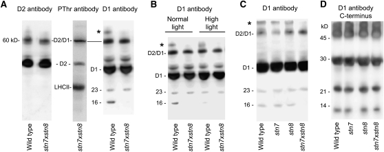
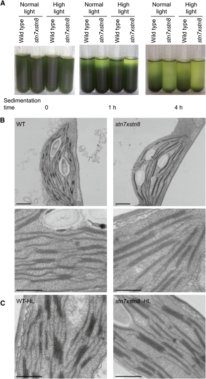
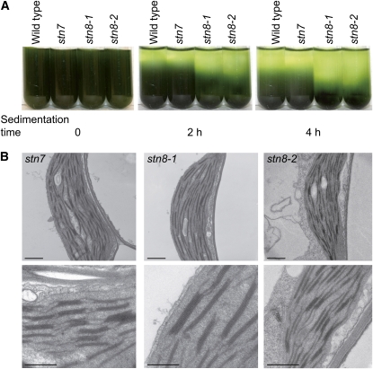
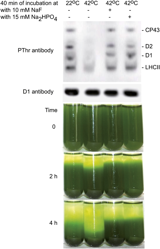
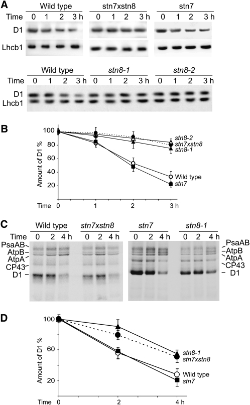
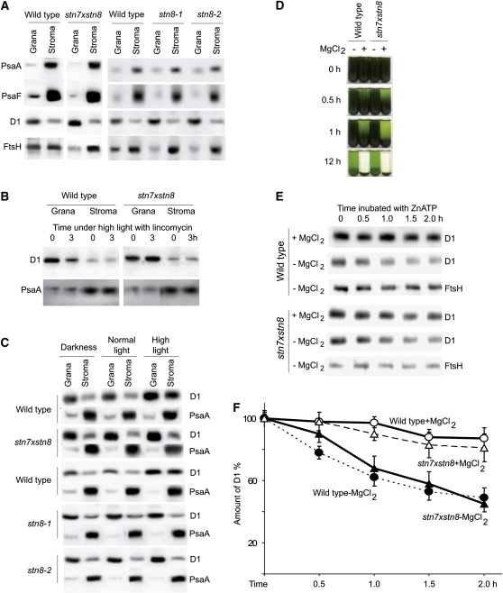
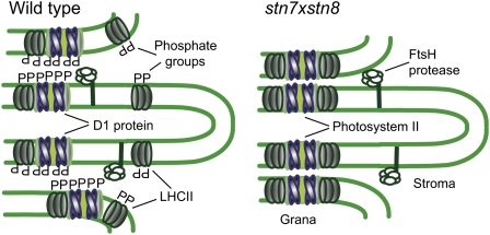
Photosynthetic thylakoid membranes in plants contain highly folded membrane layers enriched in photosystem II, which uses light energy to oxidize water and produce oxygen. The sunlight also causes quantitative phosphorylation of major photosystem II proteins. Analysis of the Arabidopsis thaliana stn7xstn8 double mutant deficient in thylakoid protein kinases STN7 and STN8 revealed light-independent phosphorylation of PsbH protein and greatly reduced N-terminal phosphorylation of D2 protein. The stn7xstn8 and stn8 mutants deficient in light-induced phosphorylation of photosystem II had increased thylakoid membrane folding compared with wild-type and stn7 plants. Significant enhancement in the size of stacked thylakoid membranes in stn7xstn8 and stn8 accelerated gravity-driven sedimentation of isolated thylakoids and was observed directly in plant leaves by transmission electron microscopy. Increased membrane folding, caused by the loss of light-induced protein phosphorylation, obstructed lateral migration of the photosystem II reaction center protein D1 and of processing protease FtsH between the stacked and unstacked membrane domains, suppressing turnover of damaged D1 in the leaves exposed to high light. These findings show that the high level of photosystem II phosphorylation in plants is required for adjustment of macroscopic folding of large photosynthetic membranes modulating lateral mobility of membrane proteins and sustained photosynthetic activity.


Figures

Figure 1.
Figure 1.
Phosphorylation of Thylakoid Proteins in stn7xstn8. (A) Immunoblotting analysis of SDS-PAGE separated thylakoid proteins from wild-type and stn7xstn8 plants with antiphosphothreonine antibody from Zymed Laboratories before (−) and after (+) the treatment of thylakoids with trypsin. (B) The product ion MS/MS spectrum obtained by collision-induced fragmentation of a singly charged phosphopeptide with m/z = 738.4 (indicated) corresponding to the N terminus of D2 protein from stn7xstn8. The ion indicated at m/z = 604.4 corresponds to the peptide that underwent a neutral loss of phosphoric acid with a mass 98. The fragment ions b (N-terminal) and y (C-terminal) are marked below and above the peptide sequence shown with the phosphorylated and acetylated Thr indicated by the lowercase t and Ac. All b ions in the spectrum are marked with an asterisk because they lost the phosphoric acid. (C) The product ion MS/MS spectrum obtained by electron transfer dissociation of the doubly charged phosphopeptide with m/z = 608.8 (indicated) corresponding to the N terminus of PsbH protein from stn7xstn8. The z fragment ions are labeled in the spectrum. Note that the mass increment between z8 and z9 is 181 D, which corresponds to the intact phosphorylated Thr residue at the second position from the peptide N terminus and is indicated by the lowercase t in the shown peptide sequence. (D) Immunoblot with antiphosphothreonine antibodies from New England Biolabs. Thylakoid membrane proteins from wild-type and mutant (stn7xstn8) plants exposed to the normal growth light for 3 h were separated by SDS-PAGE. The membranes containing 0.8 μg chlorophyll were loaded for the wild type, while the samples from stn7xstn8 contained 0.8, 3, 4, 5, and 6 μg chlorophyll, as indicated. (E) Immunoblot with antiphosphothreonine antibodies from New England Biolabs. The thylakoid membranes were isolated from the stn7xstn8 plants adapted to darkness for 15 h (D), exposed to normal light of 120 μmol photons m−2 s−1 for 3 h (NL), or to high light of 900 μmol photons m−2 s−1 for 3 h (HL). The membranes containing 2 μg chlorophyll were loaded in each lane. The positions of the major phosphorylated proteins and of the molecular markers are indicated in (A), (D), and (E).
Figure 2.
Figure 2.
Specific Pattern of the D1 Protein Aggregation and Degradation in stn7xstn8. (A) Immunoblot analysis of thylakoid membrane proteins from wild-type and stn7xstn8 plants with antibodies against D2, D1, and phosphothreonine (Zymed Laboratories), as indicated. The positions of the 60-kD aggregate of the D2 and D1 proteins (D2/D1), 23- and 16-kD fragments of the D1 protein, and of the D1 aggregate with the high molecular mass (asterisk) are indicated. (B) Immunoblot analyses of the D1 protein in stn7xstn8 compared with wild-type plants exposed to normal light (120 μmol photons m−2 s−1) or high light (900 μmol photons m−2 s−1). The sample loading corresponding to 0.6 μg of chlorophyll was used. (C) Immunoblot analyses of the D1 protein in stn7xstn8 compared with stn7, stn8, and wild-type plants exposed to normal light (120 μmol photons m−2 s−1). (D) Immunoblot analysis of thylakoid membrane proteins from wild-type, stn7, stn8, and stn7xstn8 plants with antibodies against the C terminus of the D1 protein. The positions of the molecular markers are indicated.
Figure 3.
Figure 3.
Enhanced Thylakoid Folding in stn7xstn8. (A) Time dependence of the gravity-driven sedimentation of thylakoid membranes isolated from the wild type and stn7xstn8. The thylakoid membranes were isolated from plants exposed to normal light (120 μmol photons m−2 s−1) or high light (900 μmol photons m−2 s−1), as indicated. (B) Analysis of thylakoid membranes from the wild type and stn7xstn8 by electron microscopy. Leaves from 4-week-old wild-type and stn7xstn8 seedlings were directly fixed 3 h after the start of the light phase of the growth photoperiod and prepared for transmission electron microscopy. Chloroplast sections are shown for the wild type (left panels) and stn7xstn8 (right panels). Bars in the top and bottom panels = 1 and 0.5 μm, respectively. (C) Electron microscopy analysis of thylakoid membranes from the wild type and stn7xstn8 exposed to high light of 1000 μmol photons m−2 s−1 for 3 h. Bars = 0.5 μm. [See online article for color version of this figure.]
Figure 4.
Figure 4.
Enhanced Thylakoid Folding in stn8-1 and stn8-2. (A) Time dependence of the gravity-driven sedimentation of thylakoid membranes isolated from the wild type, stn7, stn8-1, and stn8-2. (B) Analysis of thylakoid membranes from stn7, stn8-1, and stn8-2 by electron microscopy. Leaves from 4-week-old seedlings were directly fixed 3 h after the start of the light phase of the growth photoperiod and prepared for transmission electron microscopy. Chloroplast sections are shown for stn7, stn8-1, and stn8-2, as indicated. Bars in the top and bottom panels = 1 and 0.5 μm, respectively. [See online article for color version of this figure.]
Figure 5.
Figure 5.
Dephosphorylation of Thylakoid Proteins Increases Compactness of Thylakoid Membranes from Wild-Type Plants. Thylakoid membranes isolated from the wild-type plants were resuspended in buffer without MgCl2, with or without NaF or Na2HPO4, and incubated in darkness at 22 or 42°C, as indicated. Then, the membranes were twice resuspended in the buffer with 5 mM MgCl2 to restack the thylakoids. Immunoblotting analysis of SDS-PAGE separated thylakoid proteins from the samples was done with antiphosphothreonine antibody from Zymed Laboratories and D1-specific antibody (loading control), as indicated. Time dependence of the gravity-driven sedimentation of the restacked phosphorylated and dephosphorylated thylakoid membranes is shown as well. [See online article for color version of this figure.]
Figure 6.
Figure 6.
Decreased D1 Turnover in stn7xstn8 and stn8. (A) Immunoblot analysis of thylakoid proteins from wild-type, stn7, stn8-1, stn8-2, and stn7xstn8 plants with a D1-specific antibody and a control Lhcb1-specific antibody. Thylakoids were isolated from the leaves treated with lincomycin and exposed to high light of 900 μmol photons m−2 s−1 for the indicated periods of time. (B) Time dependence of the D1 protein degradation in leaves of wild-type, stn7, stn8-1, stn8-2, and stn7xstn8 plants treated with lincomycin and exposed to high light like in (A). The values are means ± se of three independent experiments for each genotype. (C) In vivo pulse-chase experiments with chloroplast proteins of wild-type, stn7, stn8-1, and stn7xstn8 plants labeled with [35S]Met and exposed to high light of 2000 μmol photons m−2 s−1. Four-week-old plants were labeled with [35S]Met for 2 h under dim light at room temperature in the presence of cycloheximide. After a 1-h chase period in dim light, including 30 min with lincomycin, plants were exposed to high light of 2000 μmol photons m−2 s−1 for 2 and 4 h, and proteins were analyzed by SDS-PAGE and phosphor imaging. Positions of the labeled subunits psaAB of photosystem I, AtpA and AtpB subunits of ATP synthase, and CP43 and D1 proteins of PSII are indicated. (D) Time dependence of the labeled D1 protein degradation in leaves of wild-type, stn7, stn8-1, and stn7xstn8 plants subjected to in vivo pulse-chase experiments under high light as shown in (C). Amounts of labeled D1 protein were normalized relative to the sum of PsaAB, AtpA, AtpB, and CP43 bands. The values are means ± se of four independent experiments of each phenotype.
Figure 7.
Figure 7.
Segregation of D1 and FtsH Protease in stn8 and stn7xstn8 Thylakoids. (A) Immunoblot analysis of PsaA, PsaF, D1, and FtsH proteins in grana and stroma membranes fractionated by digitonin treatment and differential centrifugation of thylakoids from wild-type, stn8-1, stn8-2, and stn7xstn8 plants. Equal amounts of chlorophyll were loaded on each lane. (B) Immunoblot analysis of D1 protein degradation in grana and stroma membranes fractionated by digitonin treatment of thylakoids from leaves of wild-type and stn7xstn8 plants treated with lincomycin and exposed to high light of 900 μmol photons m−2 s−1 during 0 or 3 h, as indicated. (C) Immunoblot analysis of distribution of the D1 and PsaA proteins between the grana and stroma thylakoid membranes isolated from leaves of wild-type, stn8-1, stn8-2, and stn7xstn8 plants harvested in darkness or exposed for 3 h to normal light of 120 μmol photons m−2 s−1 or to high light of 900 μmol photons m−2 s−1. (D) Time dependence of gravity-driven sedimentation of thylakoids isolated from wild-type and stn7xstn8 plants and resuspended in buffer with 5 mM MgCl2 (+MgCl2) or without MgCl2 (−MgCl2). (E) Immunoblot analysis of D1 proteolysis in thylakoids isolated from wild-type and stn7xstn8 plants and resuspended in a buffer with 5 mM MgCl2 (+MgCl2) or without MgCl2 (−MgCl2) and supplied with 0.15 mM ZnCl2 and 2 mM ATP. Immunoblotting was done using specific antibodies against the D1 and FtsH proteins, as indicated. (F) Time-dependent proteolysis of D1 in thylakoids isolated from wild-type and stn7xstn8 plants and resuspended in buffer with 5 mM MgCl2 (+MgCl2) or without MgCl2 (−MgCl2) and supplied with 0.15 mM ZnCl2 and 2 mM ATP. The values are mean ± se of three independent experiments for each experimental condition. [See online article for color version of this figure.]
Figure 8.
Figure 8.
A Model for Macroscopic Rearrangements of Plant Thylakoid Membranes via Phosphorylation-Dependent Repulsion of Adjacent Membrane Layers. The thylakoid margins of the wild type (left) and stn7xstn8 (right) are shown. The phosphate groups contributed by the PSII core proteins in the wild type loosen the appressed membrane regions through electrostatic repulsion and facilitate lateral migration of photodamaged PSII subunits from the stacked grana thylakoids to unstacked stroma lamellae, as well as the access of FtsH to the grana regions. Absence of PSII core protein phosphorylation in the stn7xstn8 mutant results in the higher membrane stacking, extension of grana regions, and limited lateral mobility of membrane proteins between the grana and stroma membrane domains. [See online article for color version of this figure.]

Similar articles

Cited by

References

    1. Adam, Z., Rudella, A., and van Wijk, K.J. (2006). Recent advances in the study of Clp, FtsH and other proteases located in chloroplasts. Curr. Opin. Plant Biol. 9 234–240. - PubMed
    1. Albertsson, P.A., and Andreasson, E. (2004). The constant proportion of grana and stroma lamellae in plant chloroplasts. Physiol. Plant. 121 334–342. - PubMed
    1. Andersson, B., and Anderson, J.M. (1980). Lateral heterogeneity in the distribution of chlorophyll-protein complexes of the thylakoid membranes of spinach chloroplasts. Biochim. Biophys. Acta 593 427–440. - PubMed
    1. Arntzen, C.J., and Ditto, C.L. (1976). Effects of cations upon chloroplast membrane subunit interactions and excitation energy distribution. Biochim. Biophys. Acta 449 259–274. - PubMed
    1. Aro, E.M., Rokka, A., and Vener, A.V. (2004). Determination of phosphoproteins in higher plant thylakoids. Methods Mol. Biol. 274 271–286. - PubMed

Publication types