. 2010 Feb 12;285(7):4273-7.
doi: 10.1074/jbc.C109.057133. Epub 2009 Dec 22.

Identification of clusterin domain involved in NF-kappaB pathway regulation

Affiliations

Identification of clusterin domain involved in NF-kappaB pathway regulation

Abdellatif Essabbani et al. J Biol Chem. .

Abstract

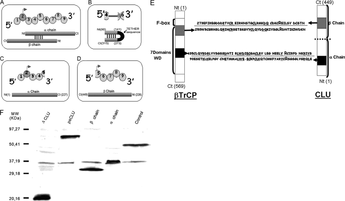
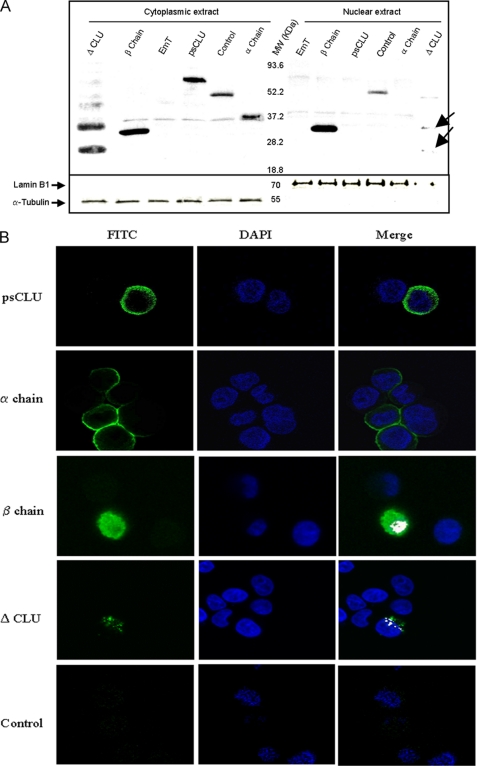
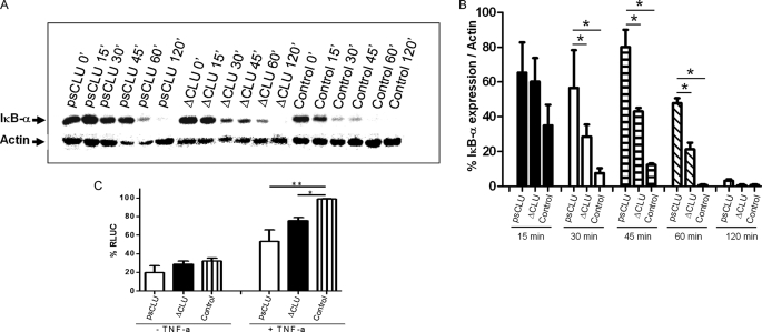
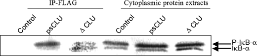
Clusterin (CLU) is a ubiquitous protein that has been implicated in tumorigenesis, apoptosis, inflammation, and cell proliferation. We and others have previously shown that CLU is an inhibitor of the NF-kappaB pathway. However, the exact form of CLU and the region(s) of CLU involved in this effect were unknown. Using newly generated molecular constructs encoding for CLU and various regions of the molecule, we demonstrated that the presecretory form of CLU (psCLU) form bears the NF-kappaB regulatory activity. Sequence comparison analysis showed sequence motif identity between CLU and beta-transducin repeat-containing protein (beta-TrCP), a main E3 ubiquitin ligase involved in IkappaB-alpha degradation. These homologies were localized in the disulfide constraint region of CLU. We generated a specific molecular construct of this region, named DeltaCLU, and showed that it has the same NF-kappaB regulatory activity as CLU. Neither the alpha-chain nor the beta-chain of CLU had any NF-kappaB regulatory activity. Furthermore, we showed that following tumor necrosis factor-alpha stimulation of transfected cells, we could co-immunoprecipitate phospho-IkappaB-alpha with DeltaCLU. Moreover, we showed that DeltaCLU could localize both in the cytoplasm and in the nucleus. These results demonstrate the identification of a new CLU activity site involved in NF-kappaB pathway regulation.


Figures

FIGURE 1.
FIGURE 1.
CLU constructs and protein expression in HeLa cells following transfection. A–D, schematic representation of psCLU, ΔCLU, α-chain, and β-chain molecular constructs, respectively (top, exonic sequence; bottom, protein structure). Ct, C terminus; Nt, N terminus. E, CLU and β-TrCP homology domains. Bold and capital letter R (for Arg) represents identified IκB-α binding sites (21). Underlined sequence letters represent homologous sequences between CLU and β-TrCP. F, Western blots of cytoplasmic protein extractions after CLU construct transfection were made with anti-FLAG antibody. pFLAG-BAP was used as a control plasmid. MW, molecular weight standards.
FIGURE 2.
FIGURE 2.
Subcellular localization of CLU constructs in HeLa cells. A, Western blot of nuclear and/or cytoplasmic protein of different CLU constructs: psCLU, ΔCLU, β-chain, and α-chain. Control and EmT correspond to pFLAG-BAP and empty vector transfection, respectively. Lamin B1 and α-tubulin were used as control for nuclear and cytoplasmic protein fractions. MW, molecular weight standards. B, confocal microscopy observation of HeLa cells transfected with psCLU-pFLAG, α-chain pFLAG, β-chain pFLAG, and ΔCLU-pFLAG. Control corresponds to nontransfected cells. The cells were incubated with mouse anti-FLAG antibody-labeled FITC (green) to detect the presence of different constructs and with DAPI (blue) to stain the nucleus. The white color corresponds to the superposition (Merge) of FITC and DAPI fluorescence.
FIGURE 3.
FIGURE 3.
ΔCLU expression delayed the degradation of IκB-α and modulated NF-κB activation. A, Western blotting of IκB-α expression at 0, 15, 30, 45, 60, and 120 min after TNF-α stimulation (5 ng/ml) in the presence of cycloheximide (100 μg/ml). Control corresponds to empty pFLAG plasmid. B, relative expression levels over time determined by densitometry as mean ± S.E. from two individual experiments. *, p value < 0.05 (Student's t test). C, HeLa cells were co-transfected with NF-κB-dependent (3E-κB-Cona luc) DNA luciferase expression vector (1 μg) and cytomegalovirus vector pFLAG (2.5 μg) expressing either full CLU or ΔCLU. Empty vectors were transfected as control. After transfection, cells were left untreated (−TNF-α) or stimulated with TNF-α (+TNF-α) for 6 h. Experiments were repeated three times. *, p value = 0.0075 (Student's t test), **, p value = 0.0498 (Mann-Whitney test). RLUC, relative luminescent units.
FIGURE 4.
FIGURE 4.
Identification of the molecular partners of psCLU and ΔCLU. The HeLa cells were transfected with control (empty vector pFLAG) and psCLU- and ΔCLU-encoding plasmids. The immunoprecipitations (IP) were carried out after inhibition of proteasome (MG132 at 20 μm for 4 h) and TNF-α stimulation (20 ng/ml) for 10 min.

Similar articles

Cited by

References

    1. Jones S. E., Jomary C. (2002) Int. J. Biochem. Cell Biol. 34, 427–431 - PubMed
    1. Trougakos I. P., Gonos E. S. (2002) Int. J. Biochem. Cell Biol. 34, 1430–1448 - PubMed
    1. Chen X., Halberg R. B., Ehrhardt W. M., Torrealba J., Dove W. F. (2003) Proc. Natl. Acad. Sci. U.S.A. 100, 9530–9535 - PMC - PubMed
    1. Leskov K. S., Klokov D. Y., Li J., Kinsella T. J., Boothman D. A. (2003) J. Biol. Chem. 278, 11590–11600 - PubMed
    1. Trougakos I. P., Djeu J. Y., Gonos E. S., Boothman D. A. (2009) Cancer Res. 69, 403–406 - PMC - PubMed

Publication types