. 2010 Feb 26;285(9):6259-64.
doi: 10.1074/jbc.M109.038570. Epub 2009 Dec 22.

c-Jun N-terminal kinase enhances MST1-mediated pro-apoptotic signaling through phosphorylation at serine 82

Affiliations

c-Jun N-terminal kinase enhances MST1-mediated pro-apoptotic signaling through phosphorylation at serine 82

Wenzhi Bi et al. J Biol Chem. .

Abstract

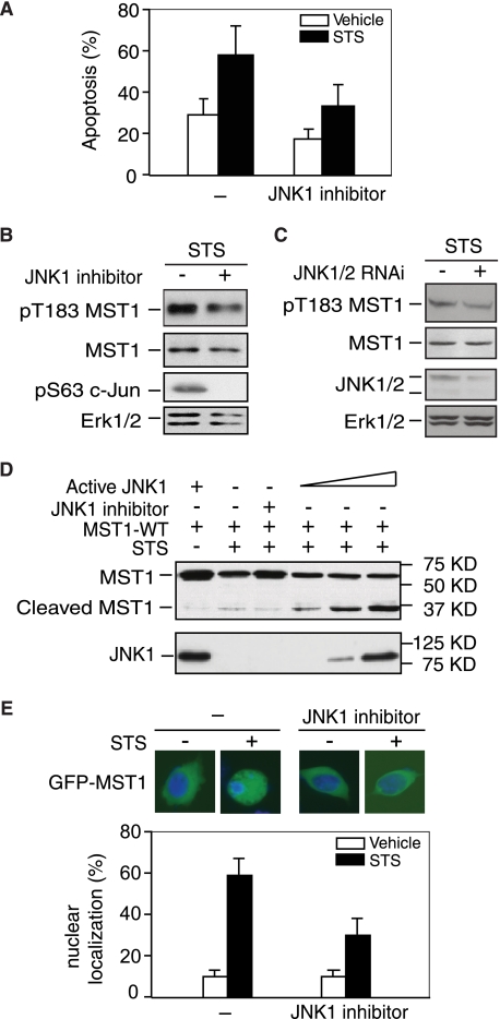
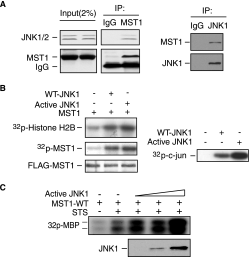
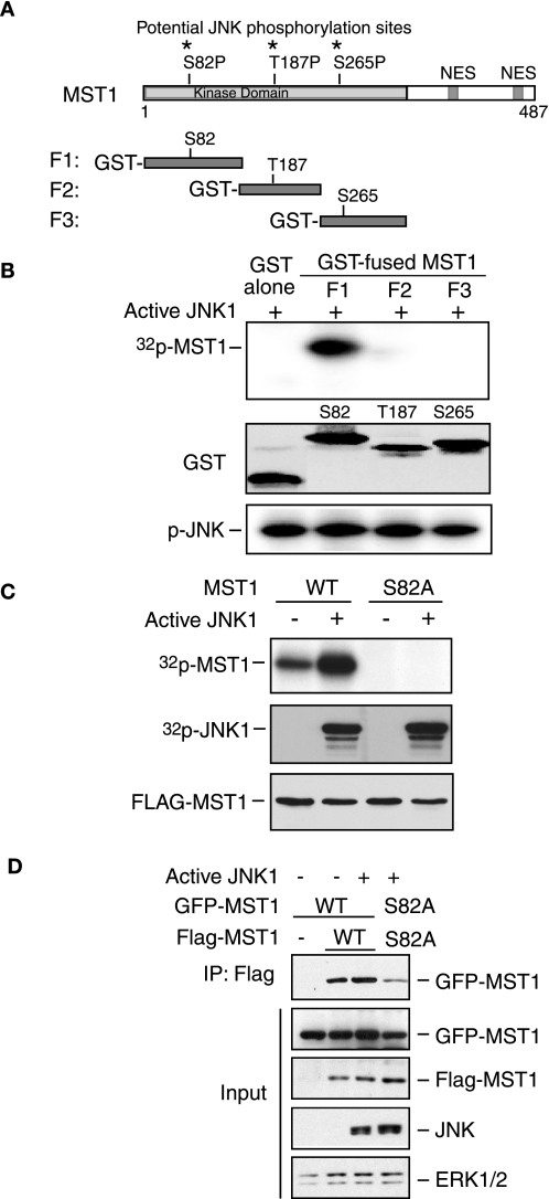
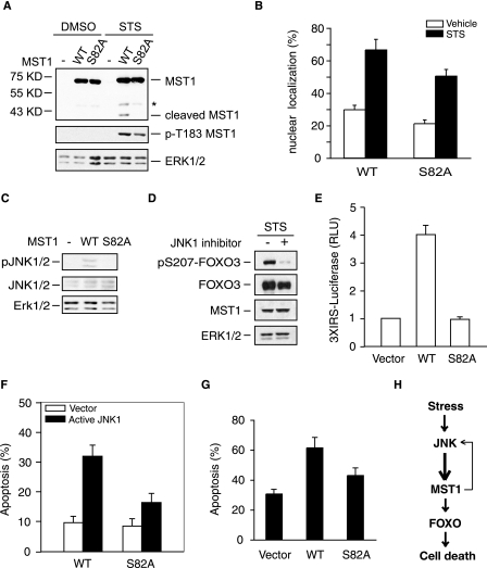
Protein kinases play an important role in the maintenance of homeostasis between cell survival and apoptosis. Deregulation of these kinases leads to various pathological manifestations, such as cancer and neurodegenerative diseases. The MST1 encodes a serine/threonine kinase that is activated upon apoptotic stimulation, which in turn phosphorylates its downstream targets, Histone H2B and FOXO. However, the upstream regulators of MST1 kinase have been poorly studied. In this study, we report that JNK (c-Jun N-terminal kinase) phosphorylates MST1 at serine 82, which leads to the enhancement of MST1 activation. Accordingly, the activation of MST1 phosphorylates FOXO3 at serine 207 and promotes cell death. The inhibition of JNK kinase per se attenuates MST1 activity and nuclear translocation as well as MST1-induced apoptosis. We also find the S82A (serine mutated to alanine) diminishes MST1 activation and its effect on the FOXO transcription activity. Collectively, these findings define the novel feedback regulation of MST1 kinase activation by its putative substrate, JNK, with implication for our understanding of the signaling mechanism during cell death.


Figures

FIGURE 1.
FIGURE 1.
JNK activity is required for MST1 activation, cleavage, and nuclear translocation. A, ovarian cancer cell line OV8 overexpressed with wild-type MST1 and treated with or without staurosporine (20 nm, 24 h) and JNK inhibitor (SP600125) and cell death was analyzed by TUNEL staining. Cell death was significantly reduced in cells treated with JNK inhibitor (n = 3, p < 0.05, ANOVA followed by Fisher's PLSD post-hoc test). B, lysates of 293T cells transfected with wild-type MST1 and treated with JNK inhibitor in the presence of STS stimulation were blotted with the pT183 MST1, FLAG, or pS63 c-Jun antibody, and Erk1/2 expression was used as the loading control. JNK inhibition reduced STS-induced MST1 autophosphorylation. C, lysates of 293T cells transfected with JNK1/2 RNAi or control vector and treated with STS (0.1 μm, 3 h) were blotted with pT183 MST1, FLAG, JNK1/2, or Erk1/2 antibody. JNK knock-down reduces MST1 activation upon STS treatment. D, lysates of COS7 cells transfected with FLAG-tagged MST1 and different amounts of active JNK1 (MKK fused JNK1) or its control vector in the presence of STS or JNK inhibitor as indicated in the figure were immunoblotted with the anti-FLAG or JNK1 antibody. Active JNK induces MST1 cleavage, and JNK inhibition decreases MST1 cleavage. E, COS7 cells transfected with GFP-MST1 and treated with STS in the absence or presence of JNK inhibitor were analyzed by fluorescence microscopy. Representative images are shown. Quantification of results is shown below, and JNK inhibitor significantly decreased GFP-MST1 nuclear translocation upon STS treatment. For each experiment, 100 cells were counted (n = 3; p < 0.01, ANOVA followed by Fisher's PLSD post-hoc test).
FIGURE 2.
FIGURE 2.
JNK interacts with MST1 and increases MST1 kinase activity. A, lysates of COS7 cells were immunoprecipitated with the anti-MST1 antibody (left panel) or anti-JNK1 antibody (right panel) or the control IgG, and were blotted with anti-JNK1/2 (left) or anti-MST1 (right) antibody. MST1 interacts with JNK. B, lysates of COS7 cells transfected with FLAG-MST1 expression plasmids together with an expression vector encoding WT-JNK1, active JNK1, or the control vector were immunoprecipitated with anti-FLAG antibody and subjected to in vitro MST1 hot kinase assay by using histone H2B as the substrate (left). MST1 autophosphorylation and expression are shown in the lower panels. JNK activity was determined by JNK kinase assay using recombinant GST-c-Jun as the substrate (right panel). JNK activation increased the MST1 activity in vitro. C, MBP in-gel kinase assay was performed using the immunoprecipitates from COS7 cells transfected with MST1 together with active JNK1 in the presence of STS stimulation. The active JNK expression level is shown in the lower panel. JNK enhances STS-induced MST1 activation.
FIGURE 3.
FIGURE 3.
JNK phosphorylates MST1 at serine 82. A, three potential JNK phosphorylation sites in the N terminus of MST1 are indicated. NES, nuclear- exporting sequence. B, in vitro JNK kinase assay was performed by incubating the recombinant active JNK with different GST-fused MST1 fragments (P1, P2, and P3) or GST only as substrate in the presence of [32P]ATP. The reaction was analyzed by SDS-PAGE followed by autoradiography. GST protein expression of MST1 and autophosphorylation of JNK are shown in the middle panel and lower panel, respectively. The phosphorylation site of MST1 by JNK kinase is in the P1 fragment. C, in vivo labeling was performed according to the “Experimental Procedures” after co-transfection with active JNK and MST1 in COS7 cells. The membrane was exposed to X-films after electrophoresis (upper panel). The expression of JNK or MST1 is shown in the lower panels. D, lysates of 293T cells transfected with GFP-MST1 (WT or S82A) and FLAG-MST1 (WT or S82A) together with an expression vector encoding active JNK1 or the control vector were immunoprecipitated with anti-FLAG antibody and blotted with GFP antibody. The input was blotted with GFP, FLAG, JNK, or Erk1/2 antibody. MST-S82A mutants confer a lower capability of dimerization.
FIGURE 4.
FIGURE 4.
Serine 82 phosphorylation is required for the activation of the MST1 signaling cascade in the cell death. A, lysates of 293T cells, transfected with FLAG-tagged WT- or S82A-MST1 and treated with or without STS, were immunoblotted with the FALG (top panel), pT183-MST1(middle panel), or Erk1/2 antibody (lower panel). Asterisk indicates the nonspecific band. The serine 82 to alanine mutation reduces the cleavage and activation of MST1. B, COS7 cells transfected with GFP-MST1 WT or S82A and treated with STS were analyzed as in Fig. 1E. S82A decreased GFP-MST1 nuclear translocation upon STS treatment (p < 0.05). C, lysates of 293T cells transfected with WT- or S82A-MST1 or the control vector were immunoblotted with the pJNK1/2 (top panel), JNK1/2 (middle panel), or Erk1/2 antibody (lower panel). The serine 82 to alanine mutation reduces the activation of JNK. D, lysates of 293T cells transfected MST1 together with FOXO3 in the treatment of JNK inhibitor or vehicle in the presence of STS were immunoblotted with the pS207-FOXO3 (top panel), GFP (FOXO3), FLAG (MST1), or Erk1/2 antibody. The inhibition of JNK kinase decreases the MST1-induced FOXO3 phosphorylation. E, cells transfected with 3XIRS luciferase plasmid and tk-Renilla reporter together with FOXO3 expression plasmid, and an expression plasmid encoding WT-MST1, S82A-MST1, or the control vector were subjected to the luciferase assay. Shown is the mean ± S.E. of the normalized fly/Renilla luciferase values relative to the control (ANOVA; p < 0.001, n = 3). The MST1-FOXO-mediated gene transcription is significantly reduced in S82A-MST1-transfected cells. F, COS7 cells were transfected with WT- or S82A-MST1 plasmid together with active JNK1 or the control vector, and cell death was analyzed as in Fig. 1A. Active JNK increases the WT-MST1, but not the S82A-MST1-induced cell death (Student's t test, p < 0.01, n = 3). G, COS7 cells were transfected with WT- or S82A-MST1 plasmid together or with the control vector in the presence of STS treatment, and cell death was analyzed as in Fig. 1A. STS induced less cell death in the S82A-MST1-transfected cells compared with WT-MST1 (p < 0.05). H, model of the JNK-MST1 signaling pathway.

References

    1. Creasy C. L., Ambrose D. M., Chernoff J. (1996) J. Biol. Chem. 271, 21049–21053 - PubMed
    1. Harvey K. F., Pfleger C. M., Hariharan I. K. (2003) Cell 114, 457–467 - PubMed
    1. Wu S., Huang J., Dong J., Pan D. (2003) Cell 114, 445–456 - PubMed
    1. Huang J., Wu S., Barrera J., Matthews K., Pan D. (2005) Cell 122, 421–434 - PubMed
    1. Taylor L. K., Wang H. C., Erikson R. L. (1996) Proc. Natl. Acad. Sci. U.S.A. 93, 10099–10104 - PMC - PubMed

Publication types