. 2010 Feb 19;285(8):5296-307.
doi: 10.1074/jbc.M109.040733. Epub 2009 Dec 22.

STAT5 activation is critical for the transformation mediated by myeloproliferative disorder-associated JAK2 V617F mutant

Affiliations

STAT5 activation is critical for the transformation mediated by myeloproliferative disorder-associated JAK2 V617F mutant

Megumi Funakoshi-Tago et al. J Biol Chem. .

Abstract

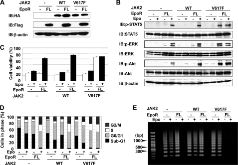
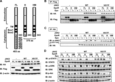
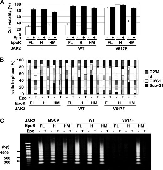
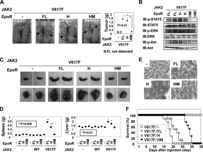
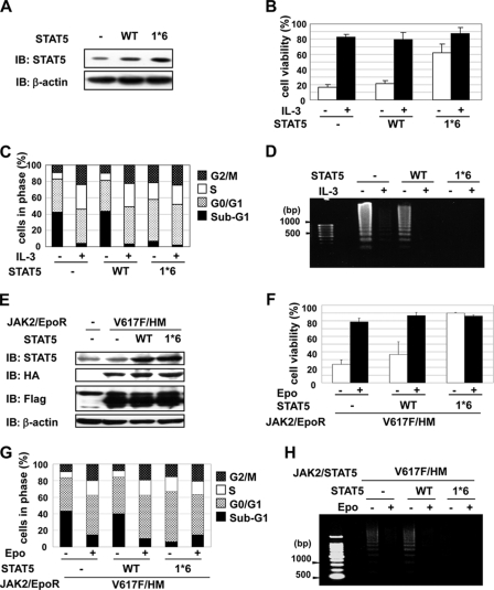
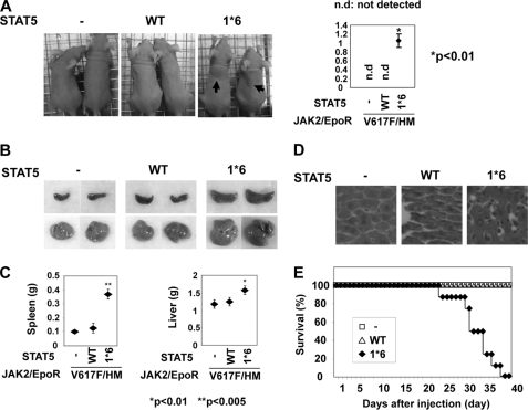
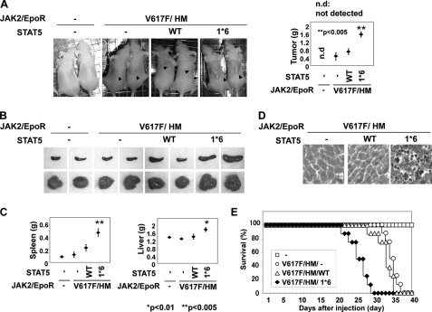
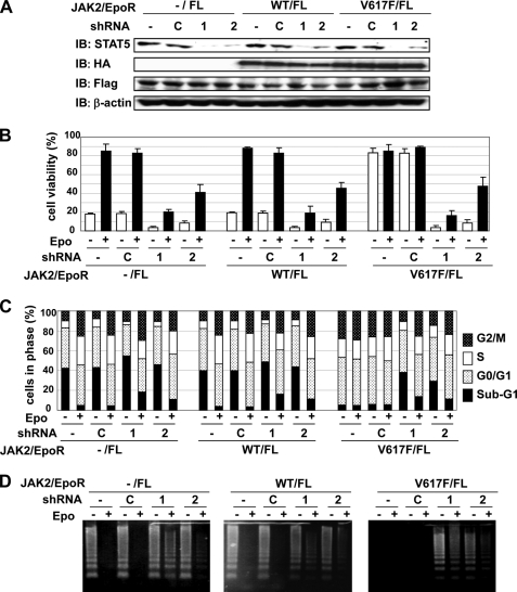
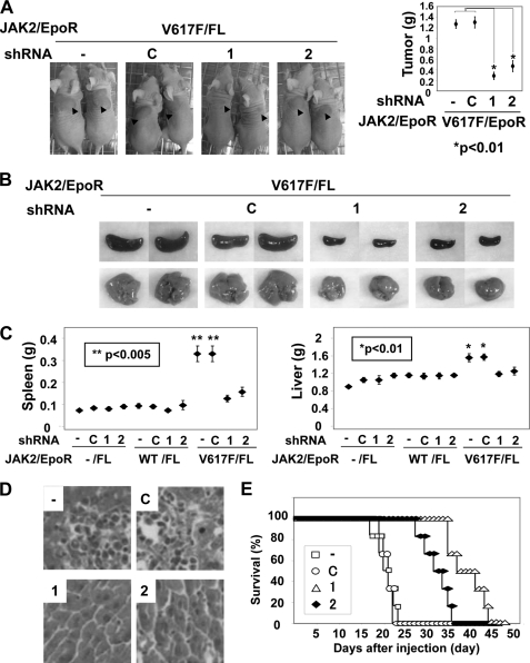
It has been well established that disruption of JAK2 signaling regulation is involved in various hematopoietic disorders; however, the detailed mechanism by which abnormal activation of JAK2 exhibits transforming activity remains to be elucidated. Here, to clarify the functional role of the erythropoietin receptor (EpoR) and its downstream transcription factor STAT5 in the abnormal activation of JAK2-induced hematopoietic diseases, we generated a stable transfectant of Ba/F3 cells expressing EpoR and analyzed the molecular mechanism of how JAK2 mutation induces cell growth disorder. JAK2 V617F mutant exhibited transforming activity when EpoR was coexpressed. According to a study utilizing several truncated mutants of EpoR, the ability of EpoR to facilitate the transforming activity of JAK2 V617F mutant required the intracellular domain to interact with STAT5. Strikingly, once the truncated EpoR (EpoR-H) was mutated on Tyr-343, the phosphorylation of which is known to be important for interaction with STAT5, JAK2 V617F mutant failed to exhibit transforming activity, suggesting that STAT5 is critical for JAK2 mutant-induced hematopoietic disorder. Furthermore, the expression of the constitutively active STAT5 mutant exhibited transforming activity in Ba/F3 cells, and short hairpin RNA-mediated knockdown of STAT5 significantly inhibited the transforming activity of JAK2 V617F mutant. Taking these observations together, STAT5 plays an essential role in EpoR-JAK2 V617F mutant-induced hematopoietic disorder. Although it remains unclear why the presence of EpoR is required to activate oncogenic signaling via the JAK2 mutant and STAT5, its interacting ability is a target for the treatment of these hematopoietic diseases.


Figures

FIGURE 1.
FIGURE 1.
EpoR is required for cytokine-independent cell survival induced by JAK2 V617F mutant. Ba/F3 cell lines were infected with empty virus (−) and retrovirus encoding wild-type JAK2 c-HA or JAK2 mutant c-HA (V617F) and EpoR c-FLAG (FL). Wild-type JAK2, JAK2 V617F mutant, and full-length EpoR are shown as WT, V617F, and FL, respectively. Each cell strain was named −/−, −/FL, WT/−, WT/FL, V617F/−, and V617F/FL cells. A, cell lysates of transduced Ba/F3 cells were blotted with anti-HA antibody, anti-FLAG antibody, or anti-β-actin antibody. B, transduced Ba/F3 cells were washed twice with PBS and left untreated or stimulated with Epo (5 units/ml) for 24 h. Whole cell lysates were immunoblotted (IB) with anti-phospho-STAT5 antibody (Tyr-694), anti-STAT5 antibody, anti-phospho-ERK antibody (Thr-202/Tyr-204), anti-ERK antibody, anti-phospho-Akt antibody (Ser-473), anti-Akt antibody, or anti-β-actin antibody. C, transduced Ba/F3 cells were washed twice with PBS and left untreated or stimulated with Epo (5 units/ml) for 48 h. The viability of these cells was determined by the trypan blue exclusion method. Results represent the mean ± S.D. of three independent experiments. D and E, transduced Ba/F3 cells were washed twice with PBS and left untreated or stimulated with Epo (5 units/ml) for 24 h. D, cells were fixed, treated with propidium iodide, and subjected to FACS analysis, as described under “Experimental Procedures.” E, DNA was isolated from cells and subjected to agarose gel electrophoresis.
FIGURE 2.
FIGURE 2.
Phosphorylation at Tyr-343 in EpoR is required for constitutive activation of STAT5 induced by JAK2 V617F mutant. A, schematic diagram of full-length EpoR and deletion mutants of EpoR (H and HM). The relative positions of tyrosine residues are marked. EpoR-HM mutant harbors Y343F substitution. TM indicates the transmembrane region, and aa indicates amino acid. JAK2 interacts with EpoR through Box1 and Box2 regions (upper panel). Ba/F3 cell lines were infected with empty virus (−) and retrovirus encoding wild-type JAK2 c-HA or JAK2 mutant c-HA (V617F) with EpoR c-FLAG (FL), EpoR-H c-FLAG (H), or EpoR-HM c-FLAG (HM). Wild-type JAK2, JAK2 V617F mutant, full-length EpoR, EpoR-H mutant, and EpoR-HM mutant are shown as WT, V617F, FL, H, and HM, respectively. Each cell strain was named −/−, −/FL, −/H, −/HM, WT/−, WT/FL, WT/H, WT/HM, V617F/−, V617F/FL, V617F/H, and V617F/HM cells. Cell lysates of transduced Ba/F3 cells were blotted with anti-HA antibody, anti-FLAG antibody, or anti-β-actin antibody (bottom). B–D, transduced Ba/F3 cells were washed twice with PBS and left untreated or stimulated with Epo (5 units/ml) for 24 h. B, cell lysates were subjected to immunoprecipitation (IP) using anti-FLAG antibody and immunoblotted (IB) with anti-HA antibody or anti-FLAG antibody. C, cell lysates were subjected to immunoprecipitation using anti-HA antibody and immunoblotted with anti-phospho-JAK2 antibody (Tyr-1007/1008) or anti-HA antibody. D, whole cell lysates were immunoblotted with anti-phospho-STAT5 antibody (Tyr-694), anti-STAT5 antibody, anti-phospho-ERK antibody (Thr-202/Tyr-204), anti-ERK antibody, anti-phospho-Akt antibody (Ser-473), anti-Akt antibody, or anti-β-actin antibody.
FIGURE 3.
FIGURE 3.
STAT5 activation is required for cytokine-independent cell survival induced by JAK2 V617F mutant. Ba/F3 cell lines were infected with empty virus (−) and retrovirus encoding wild-type JAK2 c-HA or JAK2 mutant c-HA (V617F) with EpoR c-FLAG (FL), EpoR-H c-FLAG (H), or EpoR-HM c-FLAG (HM). Wild-type JAK2, JAK2 V617F mutant, full-length EpoR, EpoR-H mutant, and EpoR-HM mutant are shown as WT, V617F, FL, H, and HM, respectively. A, transduced Ba/F3 cells were washed twice with PBS and left untreated or stimulated with Epo (5 units/ml) for 48 h. The viability of these cells was determined by the trypan blue exclusion method. Results represent the mean ± S.D. of three independent experiments. B and C, transduced Ba/F3 cells were washed twice with PBS and left untreated or stimulated with Epo (5 units/ml) for 24 h. B, cells were fixed, treated with propidium iodide, and subjected to FACS analysis, as described under “Experimental Procedures.” C, DNA was isolated from cells and subjected to agarose gel electrophoresis.
FIGURE 4.
FIGURE 4.
STAT5 activation is required for JAK2 V617F mutant-induced tumor formation in nude mice. Ba/F3 cell lines were infected with retrovirus empty virus (−) and encoding wild-type JAK2 c-HA or JAK2 mutant c-HA (V617F) with EpoR c-FLAG (FL), EpoR-H c-FLAG (H), or EpoR-HM c-FLAG (HM). Wild-type JAK2, JAK2 V617F mutant, full-length EpoR, EpoR-H mutant, and EpoR-HM mutant are shown as WT, V617F, FL, H, and HM, respectively. 1 × 10⁷ cells of transduced Ba/F3 cells were subcutaneously injected into nude mice. A, nude mice were photographed 12 days post-inoculation. Arrows indicate tumors in nude mice (left). 12 days post-inoculation, tumors at injected sites were weighed and plotted. * indicates significant difference p < 0.01 (right). N.D. indicates not detected. B, 12 days post-inoculation, mice were sacrificed, and cell lysates were prepared from tumors inoculated with V617F/FL, V617F/H, and V617F/HM cells. Whole cell lysates were immunoblotted (IB) with anti-phospho-STAT5 antibody (Tyr-694), anti-STAT5 antibody, anti-phospho-ERK antibody (Thr-202/Tyr-204), anti-ERK antibody, anti-phospho-Akt antibody (Ser-473), or anti-Akt antibody. C, 12 days post-inoculation, mice were sacrificed. In nude mice injected with Ba/F3 cells expressing JAK2 mutant c-HA (V617F) with EpoR c-FLAG (FL), EpoR-H c-FLAG (H), or EpoR-HM c-FLAG (HM), morphological changes of the spleen and liver were photographed. D, 12 days post-inoculation, four mice were sacrificed, and the spleen and liver were weighed and plotted. * and ** indicate significant differences p < 0.01 and p < 0.005, respectively. E, 12 days post-inoculation, liver sections were stained with hematoxylin-eosin (magnification ×400). F, 10 nude mice were injected for Ba/F3 cell lines expressing JAK2 mutant c-HA (V617F) with EpoR c-FLAG (FL), EpoR-H c-FLAG (H), or EpoR-HM c-FLAG (HM). For 35 days post-inoculation, mouse survival was monitored daily.
FIGURE 5.
FIGURE 5.
Constitutively active mutant of STAT5 induced cytokine-independent cell survival of Ba/F3 cells and restored cell survival of Ba/F3 cells expressing JAK2 V617F and EpoR HM mutants. A–D, Ba/F3 cells were infected with empty virus (−) and retrovirus encoding wild-type STAT5 or constitutively active mutant of STAT5 (1*6). E–F, Ba/F3 cell lines were infected with retrovirus encoding JAK2 mutant c-HA (V617F) with EpoR c-FLAG (FL) and sequentially infected with empty virus (−) and retrovirus encoding wild-type STAT5 or constitutively active mutant of STAT5 (STAT5 1*6). A and E, cell lysates of transduced Ba/F3 cells were blotted with anti-STAT5 antibody, anti-HA antibody, anti-FLAG antibody, or anti-β-actin antibody. B and F, transduced Ba/F3 cells were washed twice with PBS and left untreated or stimulated with Epo (5 units/ml) for 48 h. The viability of these cells was determined by the trypan blue exclusion method. Results represent the mean ± S.D. of three independent experiments. C, D, G, and H, transduced Ba/F3 cells were washed twice with PBS and left untreated or stimulated with Epo (5 units/ml) for 24 h. C and G, cells were fixed, treated with propidium iodide, and subjected to FACS analysis as described under “Experimental Procedures.” D and H, DNA was isolated from cells and subjected to agarose gel electrophoresis. IB, immunoblot.
FIGURE 6.
FIGURE 6.
Constitutively active mutant of STAT5 induced tumor formation in nude mice. Ba/F3 cells were infected with empty virus (−) and retrovirus encoding wild-type STAT5 or constitutively active mutant of STAT5 (1*6). 1 × 10⁷ cells of transduced Ba/F3 cells were subcutaneously injected into nude mice. A, nude mice were photographed 17 days post-inoculation. Arrows indicate tumors in nude mice (left). 17 days post-inoculation, tumors at the injected sites were weighed and plotted (right). * indicates significant difference p < 0.01. n.d. indicates not detected. B, 17 days post-inoculation, mice were sacrificed. Morphological changes of the spleen and liver were photographed. C, 17 days post-inoculation, four mice were sacrificed, and the spleen and liver were weighed and plotted. * and ** indicate significant differences of p < 0.01 and p < 0.005, respectively. D, 17 days post-inoculation, liver sections were stained with hematoxylin-eosin (magnification ×400). E, eight nude mice were injected with Ba/F3 cell lines infected with empty virus (−) and retrovirus encoding wild-type STAT5 or constitutively active mutant of STAT5. For 40 days post-inoculation, mouse survival was monitored daily.
FIGURE 7.
FIGURE 7.
Constitutively active mutant of STAT5 induced tumor formation in nude mice injected with Ba/F3 cells expressing JAK2 V617F and EpoR HM mutants. Ba/F3 cell lines were infected with retrovirus encoding JAK2 mutant c-HA (V617F) with EpoR c-FLAG (FL) and sequentially infected with empty virus (−) and retrovirus encoding wild-type STAT5 or constitutively active mutant of STAT5 (1*6). 1 × 10⁷ cells of transduced Ba/F3 cells were subcutaneously injected into nude mice. A, nude mice were photographed 17 days post-inoculation. Arrowheads indicate tumors in nude mice (right). 17 days post-inoculation, tumors at the injected sites were weighed and plotted (left). * indicates significant difference; p < 0.01. n.d. indicates not detected. B, 17 days post-inoculation, mice were sacrificed. Morphological changes of the spleen and liver were photographed. C, 17 days post-inoculation, four mice were sacrificed, and the spleen and liver were weighed and plotted. * and ** indicate significant differences of p < 0.01 and p < 0.005, respectively. D, 17 days post-inoculation, liver sections were stained with hematoxylin-eosin (magnification ×400). E, eight nude mice were injected with Ba/F3 cell lines infected with retrovirus encoding JAK2 mutant c-HA (V617F), EpoR c-FLAG (HM), and empty virus (−), wild-type STAT5 or a constitutively active mutant of STAT5. For 40 days post-inoculation, mouse survival was monitored daily.
FIGURE 8.
FIGURE 8.
Knockdown of STAT5 significantly inhibited cytokine-independent cell survival induced by JAK2 V617F mutant and EpoR. Ba/F3 cell lines were infected with empty virus (−) and retrovirus encoding wild-type JAK2 c-HA or JAK2 mutant c-HA (V617F) with EpoR c-FLAG (FL). Transduced BaF3 cells were sequentially infected with retrovirus harboring control shRNA or two kinds of shRNAs against murine STAT5 (panels 1 and 2). After puromycin selection, cells were harvested. A, whole cell lysates were immunoblotted (IB) with anti-STAT5 antibody, anti-HA antibody, anti-FLAG antibody, or anti-β-actin antibody. C, control. B, transduced Ba/F3 cell lines with shRNAs were washed twice with PBS and left untreated or stimulated with Epo (5 units/ml) for 24 h. The viability of these cells was determined by the trypan blue exclusion method. Results represent the mean ± S.D. of three independent experiments. C, cells were fixed, treated with propidium iodide, and subjected to FACS analysis, as described under “Experimental Procedures.” D, transduced Ba/F3 cells were washed twice with PBS and left untreated or stimulated with Epo (5 units/ml) for 24 h. DNA was isolated from cells and subjected to agarose gel electrophoresis.
FIGURE 9.
FIGURE 9.
Knockdown of STAT5 significantly inhibited tumor formation in nude mice induced by Ba/F3 cells expressing JAK2 V617F and EpoR. Ba/F3 cell lines were infected with empty virus (−) and retrovirus encoding wild-type JAK2 c-HA or JAK2 mutant c-HA (V617F) with EpoR c-FLAG (FL). Transduced BaF3 cells were sequentially infected with retrovirus harboring control (C) shRNA and two kinds of shRNAs against murine STAT5 (panels 1 and 2). After puromycin selection, 1 × 10⁷ cells of transduced Ba/F3 cells were subcutaneously injected into nude mice. A, nude mice inoculated with Ba/F3 cells expressing JAK2 V617F and EpoR (V617F/FL) with control shRNA or shRNA against murine STAT5 (panels 1 and 2) were photographed 17 day post-inoculation. Arrowheads indicate tumors in nude mice (left). 17 days post-inoculation, tumors at injected sites were weighed and plotted. * indicates significant difference p < 0.01 (right). B, 17 days post-inoculation, the mice were sacrificed. In nude mice injected with Ba/F3 cells expressing JAK2 mutant c-HA (V617F) and EpoR c-FLAG (FL) with control shRNA or shRNA against murine STAT5 (panels 1 and 2), morphological changes of the spleen and liver were photographed. C, 17 days post-inoculation, four mice were sacrificed, and the spleen and liver were weighed and plotted. * and ** indicate significant differences of p < 0.01 and p < 0.005, respectively. D, 17 days post-inoculation, liver sections were stained with hematoxylin-eosin (magnification ×400). E, eight nude mice were injected for Ba/F3 cell lines expressing JAK2 mutant c-HA (V617F) with EpoR c-FLAG (FL) with control shRNA or shRNA against murine STAT5 (panels 1 and 2). For 50 days post-inoculation, mouse survival was monitored daily.

References

    1. Ihle J. N., Gilliland D. G. (2007) Curr. Opin. Genet. Dev. 17, 8–14 - PubMed
    1. Parganas E., Wang D., Stravopodis D., Topham D. J., Marine J. C., Teglund S., Vanin E. F., Bodner S., Colamonici O. R., van Deursen J. M., Grosveld G., Ihle J. N. (1998) Cell 93, 385–395 - PubMed
    1. Neubauer H., Cumano A., Müller M., Wu H., Huffstadt U., Pfeffer K. (1998) Cell 93, 397–409 - PubMed
    1. Um M., Lodish H. F. (2006) J. Biol. Chem. 281, 5648–5656 - PubMed
    1. Minoo P., Zadeh M. M., Rottapel R., Lebrun J. J., Ali S. (2004) Blood 103, 1398–1407 - PubMed

Publication types

MeSH terms