. 2010 Feb 19;285(8):5726-37.
doi: 10.1074/jbc.M109.046649. Epub 2009 Dec 22.

Interaction between Hhex and SOX13 modulates Wnt/TCF activity

Affiliations

Interaction between Hhex and SOX13 modulates Wnt/TCF activity

Vanessa Marfil et al. J Biol Chem. .

Abstract

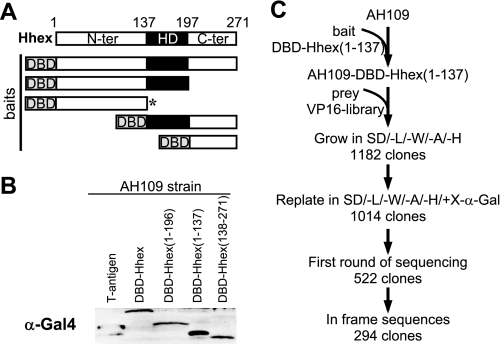
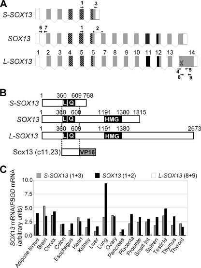
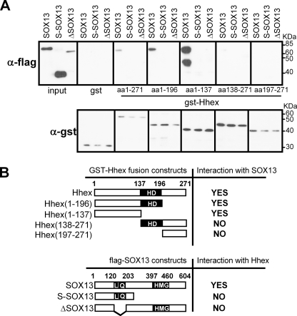
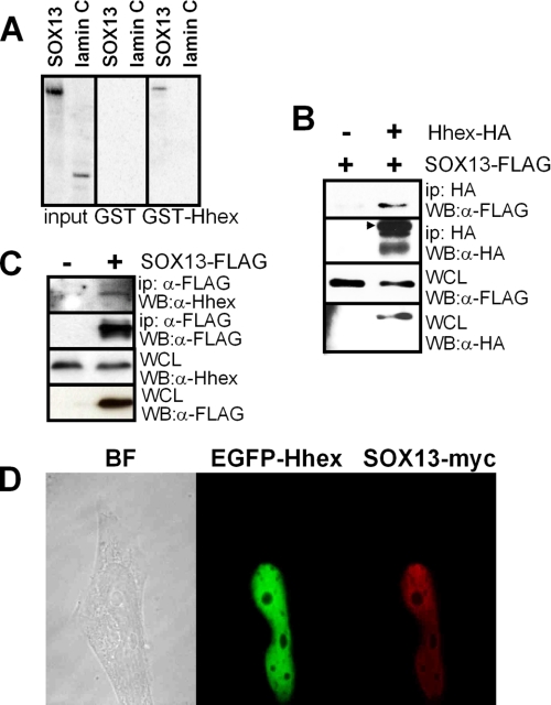
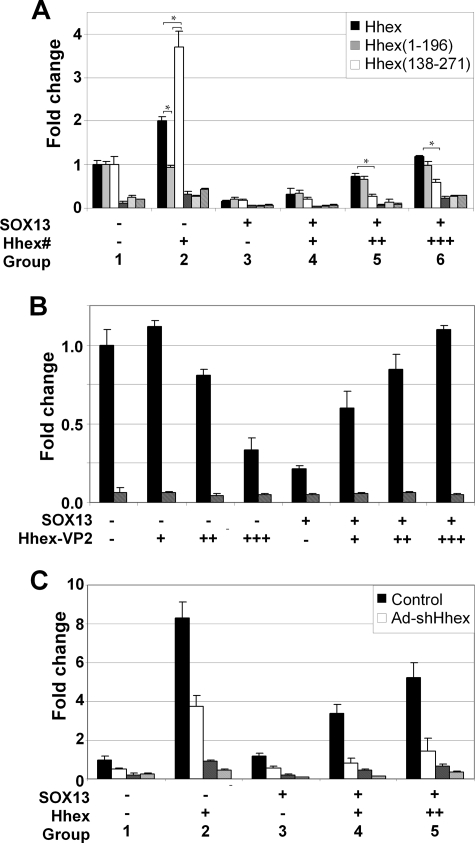
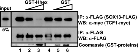
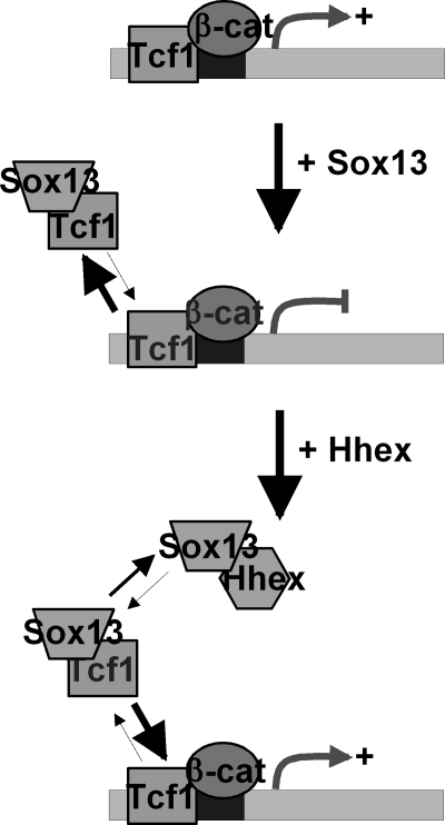
Fine-tuning of the Wnt/TCF pathway is crucial for multiple embryological processes, including liver development. Here we describe how the interaction between Hhex (hematopoietically expressed homeobox) and SOX13 (SRY-related high mobility group box transcription factor 13), modulates Wnt/TCF pathway activity. Hhex is a homeodomain factor expressed in multiple endoderm-derived tissues, like the liver, where it is essential for proper development. The pleiotropic expression of Hhex during embryonic development and its dual role as a transcriptional repressor and activator suggest the presence of different tissue-specific partners capable of modulating its activity and function. While searching for developmentally regulated Hhex partners, we set up a yeast two-hybrid screening using an E9.5-10.5 mouse embryo library and the N-terminal domain of Hhex as bait. Among the putative protein interactors, we selected SOX13 for further characterization. We found that SOX13 interacts directly with full-length Hhex, and we delineated the interaction domains within the two proteins. SOX13 is known to repress Wnt/TCF signaling by interacting with TCF1. We show that Hhex is able to block the SOX13-dependent repression of Wnt/TCF activity by displacing SOX13 from the SOX13 x TCF1 complex. Moreover, Hhex de-repressed the Wnt/TCF pathway in the ventral foregut endoderm of cultured mouse embryos electroporated with a SOX13-expressing plasmid. We conclude that the interaction between Hhex and SOX13 may contribute to control Wnt/TCF signaling in the early embryo.


Figures

FIGURE 1.
FIGURE 1.
Hhex yeast two-hybrid screening. A, a diagram is shown of Hhex fusion proteins containing the Gal4 DNA binding domain (DBD) in the N terminus. These proteins were tested as putative bait in a yeast two-hybrid assay. An asterisk indicates the protein used as the final bait in the screening. The Hhex protein showing functional domains is also shown above as a reference. N-ter, N-terminal domain; HD, homeodomain; C-ter, C-terminal domain. B, shown is immunoblotting using an anti-Gal4 antibody of extracts from the AH109 yeast transformed with the bait. The yeast-expressing plasmid, pGBKT7-T, expressing the SV40 large antigen T protein was used as a positive control. C, an overview is shown of the Hhex yeast two-hybrid screening by sequential transformation of bait and prey (VP16 library). VP16 library, E9.5-E10.5 mouse embryo library made in the pVP16; SD/L/W/A/H, synthetic dropout medium lacking leucine, tryptophan, adenine, and histidine; SD/L/W/A/H/+X-α-gal, synthetic dropout medium lacking leucine, tryptophan, adenine, and histidine, and containing 20 μg/ml of X-α-gal.
FIGURE 2.
FIGURE 2.
Interaction between Hhex and SOX13 in yeast. A, shown is multiple alignment between mouse Sox13 and the prey clones isolated in the yeast two-hybrid screening. Relevant functional domains in Sox13, the leucine zipper (LZ), and Q-rich are emphasized. Asterisks indicate the position of leucines within the LZ domain. Consensus sequences are in red. B, a representative Sox13 prey plasmid (clone 11.23) was re-transformed into AH109 yeast expressing Hhex bait or empty plasmid as a negative control. The transformed yeast were plated in SD/−Leu/−Trp and SD/−Leu/−Trp/−Ala/−His/+X-α-gal. C, β-galactosidase activity was also measured in yeast cotransformed with each Hhex bait and Sox13 prey plasmid (clone 11.23).
FIGURE 3.
FIGURE 3.
SOX13 isoforms present in human tissues. A, shown is a graphic representation of SOX13 exons as described in ECRBrowser (38). Exons are depicted as boxes. Exon size is not proportional to box size. Gray boxes represent translated exons. The HMG box is represented in black (exons 11–12). Dashed boxes represent the LZ-Q domain (exons 4–6). The conflictive G1950 in exon 14 is also shown. The primers used for qRT-PCR analysis are depicted with a number (1–9). Primer sequences are shown in Table 1. B, alignment of the SOX13 coding sequences are compared with the prey clone 11.23 isolated from the yeast two-hybrid screening. This fragment contains the LZ-Q domain of Sox13. Numbers show base pair positions. C, relative SOX13 mRNA levels in 18 human tissues were determined by qRT-PCR. The numbers in parentheses represent the primers used for the qPCR reaction (see panel A). PBGD, porphobilinogen deaminase.
FIGURE 4.
FIGURE 4.
Hhex and SOX13 interaction domains. A, shown is an in vitro binding assay between purified GST, GST-Hhex-(1–271), GST-Hhex-(1–196), GST-Hhex-(1–137), GST-Hhex-(138–271), or GST- Hhex-(197–271) from an E. coli lysate and HeLa cell extract expressing FLAG-tagged SOX13, S-SOX13, or ΔSOX13. HeLa total protein extracts expressing SOX13-related proteins were incubated with GST-Hhex proteins. Glutathione-Sepharose was used to pull down the GST-Hhex fusion proteins, and anti-FLAG antibody was used in Western blots. One-twentieth of the extract was loaded as the input. The lower band in the SOX13 incubation was routinely observed after overnight incubation and is a C-terminal degradation product of SOX13. Lower panel, shown is an immunoblot using anti-GST antibodies in 1% of the beads. B, shown are diagrams summarizing the results obtained in the GST pulldown assays. Numbers show amino acid positions. HD, homeodomain; L, leucine zipper; Q, glutamine-rich; HMG, high mobility group domain.
FIGURE 5.
FIGURE 5.
Interaction nature and co-localization of Hhex and SOX13. A, 35S-SOX13 and 35S-lamin C (negative control) were synthesized in an in vitro transcription and translation system and incubated with glutathione-Sepharose beads coated with GST-Hhex or GST. Bound proteins were run in SDS/PAGE and visualized using phosphorimaging. B, immunoblot (WB) analysis of 293T cell lysates transfected with SOX13-FLAG and Hhex-HA detects SOX13-FLAG after immunoprecipitation (ip) with anti-HA antibody and immunoblotting against the FLAG tag. The whole cell lysate (WCL) was immunoblotted using anti-FLAG or anti-HA to show the expression levels of SOX13-FLAG and Hhex-HA in the assay. C, immunoblot analysis of HepG2 cell lysates transfected with SOX13-FLAG detects endogenous Hhex after immunoprecipitation with anti-FLAG antibody and immunoblotting against a primary antibody against Hhex. The whole cell lysate was immunoblotted using anti-FLAG or anti-Hhex to show the expression levels of SOX13-FLAG and Hhex in the assay. D, co-localization of ectopically expressed Hhex and SOX13 is shown. HeLa cells were co-transfected with plasmids expressing the fusion protein EGFP-Hhex (green) and a SOX13-myc construct and labeled with anti-Myc antibody (red). Images were acquired with a Leica TCS-SP2 confocal microscope. BF, bright field.
FIGURE 6.
FIGURE 6.
Effect of Hhex and SOX13 interaction on Wnt activity. A, shown are functional domains of Hhex involved in Wnt regulation. HEK 293T cells were transfected with the Wnt reporter plasmid TOPflash (filled bars) and the mutated version, FOPflash (dashed bars), as a negative control. An expression vector containing activated β-catenin-S37Y was cotransfected in each condition. Those plasmids expressing SOX13 or Hhex derivatives were transfected as indicated below the graph. The means and S.E. of the relative firefly/renilla ratios and -fold change over the control of at least three experiments are shown. The total firefly/renilla ratio of untreated cells transfected with activated β-catenin (S37Y) was set as 1.0. Hhex#, Hhex-(1–271), Hhex-(1–196), or Hhex-(138–271). B, shown is dose-dependent regulation of Wnt by HhexVP2, a Hhex fusion protein containing two copies of the VP16 activator domain that converts Hhex into a transcriptional activator with or without SOX13 modulation. The same experiment settings as in panel A were used. C, Wnt activity is shown in HepG2 cells after knocking down endogenous Hhex by adenovirus-mediated expression of shRNA (Ad-shHhex). An adenovirus expressing shRNA against luciferase was used in HepG2 control cells. A plasmid expressing Hhex is represented at the bottom of the panel as Hhex. The same experiment settings as in panel A were used. *, p < 0.05, paired Student's t test.
FIGURE 7.
FIGURE 7.
Hhex and SOX13 competitive immunoprecipitation assay. A lysate from 293T cells co-transfected with SOX13-FLAG- and TCF1-myc-tagged-expressing plasmids was immunoprecipitated (IP) using anti-FLAG antibody in the absence of GST proteins (lanes 1 and 4) or with increasing amounts of purified GST-Hhex (lanes 2 and 3) or GST (lanes 5 and 6). The immunoprecipitated material was immunoblotted (WB) with anti-Myc antibody. The presence of increasing amounts of GST-proteins is shown with Coomassie staining of a gel run in parallel.
FIGURE 8.
FIGURE 8.
Wnt activity in mouse embryos electroporated with SOX13 and Hhex. A, a diagram shows the orientation of the embryo in the electroporation cuvette. E8.5 mouse embryos were orientated with a 45° angle to specifically transfer DNA to the pre-hepatic and pre-pancreatic endoderm. The region targeted by DNA is highlighted in red. After electroporation, embryos were incubated for 24 h in vitro. Turned-embryos, with the E9.5 developmental stage, were then dissected to isolate the liver bud region, and the reporter activity was measured. B, luciferase activity is shown of isolated liver buds from non-electroporated (first chart) or electroporated embryos with TOPflash reporter, SOX13, and/or Hhex. The total firefly/renilla ratio of embryos electroporated with TOPflash alone was set at 1.0. Each condition was done at least three times, and the total number of embryos recovered and assayed was at least six.
FIGURE 9.
FIGURE 9.
Regulation of DKK1, a target of the β-catenin-TCF pathway, by Hhex. A, shown is a schematic representation of the human DKK1 gene. TCF binding sites are depicted as black boxes. B, 293T cells were transfected with plasmids expressing TCF1-myc, SOX13, or Hhex, as shown below the graph. The expression of DKK1 was assessed by quantitative RT-PCR. C, shown is immunoblot analysis of the transfected 293T cells. D, 293T cells transfected as in B were cross-linked and lysed. The relative amounts of TCF1-myc on DKK1 promoter were analyzed by real-time qPCR of immunoprecipitated chromatin using anti-Myc antibody. Binding of TCF1 to the RPLP0 gene was used as a negative control. The image of a representative qPCR stopped at the exponential phase is also shown. Data are the mean of three experiments. PBGD, porphobilinogen deaminase.
FIGURE 10.
FIGURE 10.
Model depicting the role of Hhex in Wnt activity modulation. In this model Wnt-responsive genes are up-regulated upon the binding of a complex containing TCF1-β-catenin to consensus TCF1 binding sites. Wnt activity is inhibited in the presence of SOX13 because of its interaction with the TCF1 protein, which disrupts the TCF1-β-catenin complex. The addition of Hhex results in the formation of a Hhex·SOX13 complex, displacing SOX13 from TCF1 and allowing TCF1 to form the effector complex in sensitive promoters once more.

Similar articles

Cited by

References

    1. McLin V. A., Rankin S. A., Zorn A. M. (2007) Development 134, 2207–2217 - PubMed
    1. Ober E. A., Verkade H., Field H. A., Stainier D. Y. (2006) Nature 442, 688–691 - PubMed
    1. Thomas P. Q., Brown A., Beddington R. S. (1998) Development 125, 85–94 - PubMed
    1. Bogue C. W., Zhang P. X., McGrath J., Jacobs H. C., Fuleihan R. L. (2003) Proc. Natl. Acad. Sci. U.S.A. 100, 556–561 - PMC - PubMed
    1. Bort R., Martinez-Barbera J. P., Beddington R. S., Zaret K. S. (2004) Development 131, 797–806 - PubMed

Publication types

MeSH terms