. 2009 Dec 22;2(102):ra86.
doi: 10.1126/scisignal.2000217.

Cbl controls EGFR fate by regulating early endosome fusion

Affiliations

Cbl controls EGFR fate by regulating early endosome fusion

Gina D Visser Smit et al. Sci Signal. .

Abstract

Amino acid residues 1 to 434 of the E3 ubiquitin ligase Cbl control signaling of the epidermal growth factor receptor (EGFR) by enhancing its ubiquitination, down-regulation, and lysosomal degradation. This region of Cbl comprises a tyrosine kinase-binding domain, a linker region, a really interesting new gene finger (RF), and a subset of the residues of the RF tail. In experiments with full-length alanine substitution mutants, we demonstrated that the RF tail of Cbl regulated biochemically distinct checkpoints in the endocytosis of EGFR. The Cbl- and ubiquitin-dependent degradation of the regulator of internalization hSprouty2 was compromised by the Val(431)--> Ala mutation, whereas the Cbl- and EGFR-dependent dephosphorylation or degradation of the endosomal trafficking regulator Hrs was compromised by the Phe(434)--> Ala mutation. Deregulated phosphorylation of Hrs correlated with inhibition of the fusion of early endosomes and of the degradation of EGFR. This study provides the first evidence that Cbl regulates receptor fate by controlling the fusion of sorting endosomes. We postulate that it does so by modulating the abundance of tyrosine-phosphorylated Hrs.


Figures

Figure 1
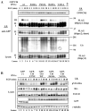
Figure 1. Biochemical characterization of Cbl RF tail alanine substitution mutants
A. The conserved C-terminal flank of the RING finger in human Cbl isoforms. Domains relevant to EGF-R regulation include: the tyrosine kinase-binding domain (TKB); linker region (L); RING finger domain (RF); RING finger C-terminal flank or “tail” domain (T); proline-rich region (PRO); and leucine zipper (LZ). Residue Y371 lies within the linker region. RING finger tail sequences of human Cbl isoforms (c-Cbl, Cbl-b, and Cbl-3) are aligned in the expanded portion of the graphic. Evolutionarily conserved sequences are marked with shaded (sequence identity) or plain (amino acid similarity) boxes. Underlined residues were substituted for this study. The vertical dashed line marks the C-terminal limit of the evolutionarily conserved Cbl sequences sufficient to enhance EGF-R ubiquitination, downregulation, and degradation. B. Cbl RF tail substitution mutants V431A and F434A are compromised for EGF-R downregulation. Surface EGF-R remaining at each stimulation time point was expressed relative to the amount of surface receptor in matched unstimulated cells. Results reflect the mean of three independent experiments ± S.D. C. Cbl RF tail substitution mutants V430A, V431A and F434A effect reduced EGF-R ubiquitination after 10 minutes of cell incubation with EGF. Upper section: 750 μg immunoprecipitates (anti-EGF-R antibody 528). Isotype-matched anti-Syk antibody 4D10 [C] was the specificity control. Immunoprecipitates (I.P.) and 75 μg lysate protein samples were gel-resolved and sequentially immunoblotted (I.B.). Lower section: Quantification of results. Ub signals were normalized to their matching EGF-R signals. Mean values were expressed relative to the signal achieved with GFP-Cbl wild type (1.00). Results represent data from three independent experiments +/− S.D. Using Student’s t-test with α=0.05, only V431A and V430A produced significantly less EGF-R ubiquitination than wt Cbl. D. Cbl mutants F434A and V431A effect reduced and delayed EGF-R ubiquitination and reduced EGF-R degradation. Upper section: 1 mg anti-EGF-R (528) immunoprecipitates. I.P.s (top three panels) and 100 μg of lysate protein from each sample (bottom panel) were sequentially immunoblotted using the antibodies indicated. The double claret marks the position of the predominant species of ubiquitinated EGF-R. Lower section: Quantification of EGF-R degradation effected by wild type and RF tail mutant Cbl proteins (100 μg protein/lane).
Figure 2
Figure 2. Aberrant regulation of hSprouty2 and Hrs by RF tail substitution mutants
A. Cbl mutants V431A and F434A are grossly and moderately compromised, respectively, for ubiquitin-associated degradation of the regulatory protein hSprouty2, but they are not defective in hSprouty2 binding. Immunoprecipitates (1 mg lysate protein per reaction; upper panels, I.P.) and 100 μg lysate samples (bottom panel) were gel-resolved and probed sequentially with the indicated antibodies (I.B.). I and II mark the more abundant species of hSprouty2. Both undergo rapid EGF-induced degradation in the presence of functionally wild type Cbl (bottom panel: wt, P433A and D432A). Species III may represent ligand-induced, ubiquitinated hSprouty2. The ligand-induced increase in Cbl/hSprouty2 complexes is best visualized in lanes 17-20, where expression of Cbl mutant V431A blocks the degradation of associated hSprouty2 (second panel). Equivalent masses of lysate protein contained comparable amounts of the GFP-Cbl proteins (top panel). B. Cbl RF tail mutant F434A effects Hrs tyrosine phosphorylation with kinetics like wild type Cbl, but the mutant is compromised for apparent Hrs dephosphorylation and/or degradation. For each sample, 100 μg of cell lysate protein was immunoblotted (I.B.) with the indicated antibodies. The γ-tubulin blot shows comparable protein loading in all lanes.
Figure 3
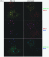
Figure 3. Cbl RF tail mutant F434A induces the aberrant pairing and clustering of early endosomes carrying activated EGF-R
COS-7 cells expressing endogenous EGF-R were transfected with the indicated GFP-Cbl expression constructs (4 μg per 10 cm dish). The cultures were stimulated with Alexa Fluor 647-EGF for 25 min, paraformaldehyde-fixed, and immunostained with anti-EEA1 antibody and a Texas Red secondary conjugate prior to visualization of the GFP-Cbl, EGF, and EEA1 signals. In five independent experiments, expression of the Cbl F434A mutant increased apparent endosome pairing and clustering. Localization of EEA1 to EGF- and GFP-Cbl-positive, clustered endosomes identifies the compartments as early endosomes. Protein colocalization within the figure is indicated by the following colors: top row, red plus green yields yellow/orange; middle row, red plus blue yields magenta; bottom row, green plus blue yields aqua/light blue (compare with the darker blue signal of the upper cell in the bottom left panel). Several examples of vesicles exhibiting protein colocalization are marked by the white arrows.
Figure 4
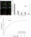
Figure 4. Deregulation of the Hrs checkpoint by Cbl mutant F434A impedes early endosome fusion
A. Select images from live cell collections (Figs. S3-S5) contrast the rapid fusion of endosomes bearing wild type GFP-Cbl with the delayed fusion of docked endosomes carrying GFP-Cbl F434A. Moderate-to-large endosomes rarely developed in Cbl V431A-expressing cells, which are shown here at reduced magnification to illustrate their distinct appearance. Note the difference in elapsed time for the paired images. For Cbl wt and F434A, Lysotracker Red-positive compartments are lysosomes. V431A images show red signal for fluor-conjugated EGF. Arrows mark the same paired vesicles at the early and later collection times. B. Fusion time was quantified from all docking events observed in movies for nine GFP-Cbl wt live cells and twelve GFP-Cbl F434A cells. Time to fusion (x-axis) is expressed relative to independent vesicle docking times, rather than from a single time point in the collection process. Standard deviations (error bars) were calculated from pooled experimental data. C. Cox proportional hazard regression analysis of fusion events for paired docked vesicles. Fusion occurred sooner in the GFP-Cbl wt-expressing cells compared to GFP-Cbl F434A expressors (p<0.0001). The graph shows the fusion distribution based on the fitted Cox model.

References

    1. Levkowitz G, Waterman H, Zamir E, Kam Z, Oved S, Langdon WY, Beguinot L, Geiger B, Yarden Y. c-Cbl/Sli-1 regulates endocytic sorting and ubiquitination of the epidermal growth factor receptor. Genes Dev. 1998;12:3663–36674. - PMC - PubMed
    1. Lill NL, Douillard P, Awwad RA, Ota S, Lupher ML, Jr., Miyake S, Meissner-Lula N, Hsu VW, Band H. The evolutionarily conserved N-terminal region of Cbl is sufficient to enhance down-regulation of the epidermal growth factor receptor. J. Biol. Chem. 2000;275:367–377. - PubMed
    1. Duan L, Miura Y, Dimri M, Majumder B, Dodge IL, Reddi AL, Ghosh A, Fernandes N, Zhou P, Mullane-Robinson K, Rao N, Donoghue S, Rogers RA, Bowtell D, Naramura M, Gu H, Band V, Band H. Cbl-mediated ubiquitinylation is required for lysosomal sorting of epidermal growth factor receptor but is dispensable for endocytosis. J. Biol. Chem. 2003;278:28950–28960. - PubMed
    1. Visser GD, Lill NL. The Cbl RING finger C-terminal flank controls epidermal growth factor receptor fate downstream of receptor ubiquitination. Exp. Cell Res. 2005;311:281–293. - PubMed
    1. Haglund K, Sigismund S, Polo S, Szymkiewicz I, Di Fiore PP, Dikic I. Multiple monoubiquitination of RTKs is sufficient for their endocytosis and degradation. Nat. Cell Biol. 2003;5:461–466. - PubMed

Substances