Review
. 2009;74(3):177-90.
doi: 10.1159/000258665. Epub 2009 Dec 21.

On the independent origins of complex brains and neurons

Affiliations
Review

On the independent origins of complex brains and neurons

Leonid L Moroz. Brain Behav Evol. 2009.

Abstract

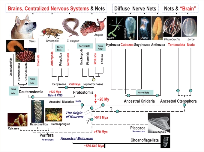
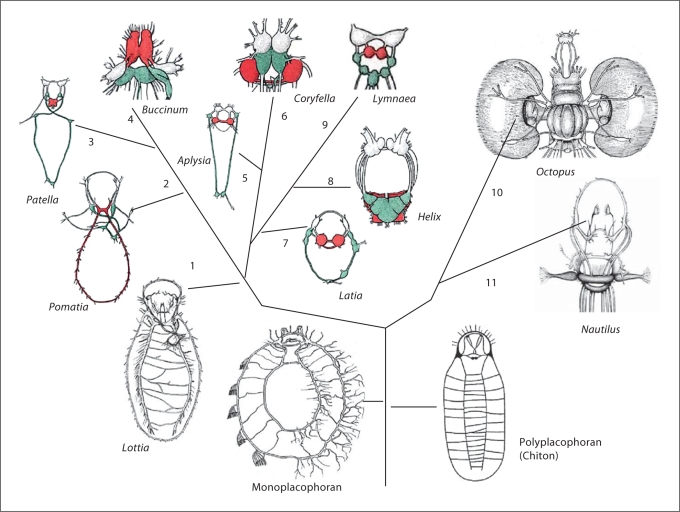
Analysis of the origin and evolution of neurons is crucial for revealing principles of organization of neural circuits with unexpected implications for genomic sciences, biomedical applications and regenerative medicine. This article presents an overview of some controversial ideas about the origin and evolution of neurons and nervous systems, focusing on the independent origin of complex brains and possible independent origins of neurons. First, earlier hypotheses related to the origin of neurons are summarized. Second, the diversity of nervous systems and convergent evolution of complex brains in relation to current views about animal phylogeny is discussed. Third, the lineages of molluscs and basal metazoans are used as illustrated examples of multiple origins of complex brains and neurons. Finally, a hypothesis about the independent origin of complex brains, centralized nervous systems and neurons is outlined. Injury-associated mechanisms leading to secretion of signal peptides (and related molecules) can be considered as evolutionary predecessors of inter-neuronal signaling and the major factors in the appearance of neurons in the first place.


Figures

Fig. 1
Fig. 1
Parallel evolution and diversity of nervous systems. The current view of evolutionary relationships in the animal kingdom is combined with the presence or absence of a central nervous system (CNS) or brain. One of the definitions of the CNS is the concentration of neuronal cells within a defined organ-type structure where neurons and neuronal processes can be supported (surrounded) by other cell types (e.g. glia or connective tissues) to maintain a controllable microenvironment for neuronal functioning. Choanoflagelates (eukaryotic algae-like organisms) are placed at the base of the tree as a sister group for Metazoa. Two basal metazoan phyla (Porifera and Placozoa) do not have recognized neurons. Two other prebilaterian/basal metazoan phyla (Cnidaria and Ctenophora) have well-defined neurons and nerves (however, only ctenophores have ‘true’ muscles of mesodermal origin). Although neuronal organization in basal Metazoa is superficially presented as a nerve net, many species have a prominent concentration of neuronal elements, and numerous and apparently autonomous networks governing surprisingly complex and well coordinated behaviors [Mackie 1990]. Cubozoa have well developed eyes and a ganglionic organization associated with rhopalia which can be described in terms of a centralized nervous system. Similarly, there is a well-defined concentration of neural elements associated with locomotory combs in Ctenophora. Chordates, nematodes, molluscs and arthropods have well-defined central nervous systems, while in other bilaterians shown in the diagram the gross anatomical organization of their nervous systems can be similar or even simpler than those in selected cnidarians and ctenophores. Centralization of nervous systems occurred in parallel within several lineages representing all three major domains in bilaterians (Deuterostomes, Ecdysozoa and Lophotrochozoa). Only representative groups of the 36 known animal phyla are shown in the diagram. Circles indicate possible events of multiple origins of neurons. See text for details. This reconstruction of phylogenetic relationships among phyla is a combined view based upon recent large-scale molecular/phylogenomic analyses of several dozen proteins from representatives of more than 15 animal phyla [Halanych, 2004; Valentine, 2004; Bourlat et al., 2006; Dunn et al., 2008; Philippe et al., 2009; Mikhailov et al., 2009]. The origin of animals can be traced back to about 600 million years ago (Mya). However, the extant animal phyla might have more recent evolutionary history. It appears that the origin of major bilaterian groups occurred within a relatively short geological time (probably within 20 million years or even less). As a result, the accurate evolutionary relationships among basal lineages and major bilaterian phyla are not well resolved (dotted lines). Possible timing of the divergence in the diagram is indicated as Mya.
Fig. 2
Fig. 2
Multiple occurrences of nervous system centralization in representatives of the phylum Mollusca. This diagram shows illustrative examples of nervous system centralization in two molluscan classes: Gastropoda (#1–9) and Cephalopoda (#10–11). The schematic outline of the anatomy of nervous systems in representatives of several lineages is combined with phylogenetic relationships among these groups. Ancestral organization of the molluscan nervous system (=tetraneury) is preserved in the two basal taxa Monoplacophora and Polyplacophora (or Chitons). Their nervous systems consist of two major elements: pedal and lateral or pleuro-parietal cords forming a visceral loop as well as cerebral and buccal loops located in the anterior part of animals. This pattern can also be recognized in Archogastropoda, represented here by the limpet Lottia (#1) and Pomatia (#2). Centralization of nervous systems occurs independently in various groups of prosobranch (#2–4), opisthobranch (#5–6), pulmonate (#7–9) and cephalopod (#10–11) molluscs. The derivatives of the pedal cords (the pedal ganglia) are shown in red; the components of the visceral loop (derivatives of pleural-parietral cords) are shown in green; and the cerebral ganglia are uncolored. Color coding has not been applied to cephalopod molluscs due to the more complex 3D organization of their brain. The modified diagrams of different molluscan nervous systems are from Bullock and Horridge [1965], fig. 22.6a, 23.3, 23.8a, 25.2, 25.5a). See text for details.

Comment in

References

    1. Arenas-Mena C, Cameron AR, Davidson EH. Spatial expression of Hox cluster genes in the ontogeny of a sea urchin. Development. 2000;127:4631–4643. - PubMed
    1. Arenas-Mena C, Martinez P, Cameron RA, Davidson EH. Expression of the Hox gene complex in the indirect development of a sea urchin. Proc Natl Acad Sci USA. 1998;95:13062–13067. - PMC - PubMed
    1. Arendt D. Genes and homology in nervous system evolution: comparing gene functions, expression patterns, and cell type molecular fingerprints. Theor Biosci. 2005;124:185–197. - PubMed
    1. Arendt D. The evolution of cell types in animals: emerging principles from molecular studies. Nat Rev Genet. 2008;9:868–882. - PubMed
    1. Arendt D, Nubler-Jung K. Inversion of dorsoventral axis? Nature. 1994;371:26. - PubMed

Publication types