Review
. 2009;74(3):219-30.
doi: 10.1159/000258668. Epub 2009 Dec 21.

Comparative neurobiology of feeding in the opisthobranch sea slug, Aplysia, and the pulmonate snail, Helisoma: evolutionary considerations

Affiliations
Review

Comparative neurobiology of feeding in the opisthobranch sea slug, Aplysia, and the pulmonate snail, Helisoma: evolutionary considerations

Margaret M Wentzell et al. Brain Behav Evol. 2009.

Abstract

The motor systems that generate feeding-related behaviors of gastropod mollusks provide exceptional opportunities for increasing our understanding of neural homologies and the evolution of neural networks. This report examines the neural control of feeding in Helisoma trivolvis, a pulmonate snail that ingests food by rasping or scraping material from the substrate, and Aplysia californica, an opisthobranch sea slug that feeds by using a grasping or seizing motion. Two classes of neurons that are present in the buccal ganglia of both species are considered: (1) clusters of peptidergic mechanoafferent cells that transmit sensory information from the tongue-like radula/odontophore complex to the central motor circuit; and (2) sets of octopamine-immunoreactive interneurons that are intrinsic to the feeding network. We review evidence that suggests homology of these cell types and propose that their roles have been largely conserved in the control of food-scraping and food-grasping consummatory behaviors. We also consider significant differences in the feeding systems of Aplysia and Helisoma that are associated with the existence of radular closure in Aplysia, an action that does not occur in Helisoma. It is hypothesized that a major adaptation in the innervation patterns of analogous, possibly homologous muscles could distinguish the food-scraping versus food-grasping species. It appears that although core CPG elements have been largely conserved in this system, the neuromuscular elements that they regulate have been more evolutionarily labile.


Figures

Fig. 1
Fig. 1
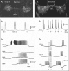
Comparative anatomy of Aplysia and Helisoma feeding systems. Left panel: the isolated odontophore-CNS preparation of Aplysia californica [modified from Rosen et al., 2000a]. This preparation includes the radula with its support structures (e.g. the rotella) and the innervation from the buccal ganglion (buccal g.) and cerebral ganglion (cerebral g.) via the radula nerve (radula n.) as well as the cerebral-buccal connective (C-B conn.). The external (dorsal) surface of the radula is viewed from above (anterior toward the top, posterior toward the bottom). This orientation corresponds to the resting position of a non-feeding specimen. During a bite, the odontophore is rotated in the anterior direction, bringing the grasping surface into contact with the potential food. During this phase of radula protraction, the two radula halves become closed around the medial longitudinal groove, serving to grasp the food prior to its subsequent retraction and backward rotation, transporting the food into the oral cavity. Right panel: A similar view of odontophore-CNS preparation of Helisoma trivolvis. Note that the lip of the odontophore at the anterior end of the radular groove is relatively broader than that of Aplysia. The odontophore of Helisoma is used as a scoop and the lateral walls do not close. The posterior buccal nerves (PB n.) of Helisoma correspond to the radula nerve of Aplysia.
Fig. 2
Fig. 2
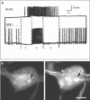
RM neurons in Aplysia and Helisoma. A1 SCP-like immunoreactive (SCP-li) neurons on the rostral surface of the Aplysia californica buccal ganglia [modified from Miller et al., 1994]. A cluster of closely apposed medium-sized (20–80 μm) sensory neurons was located in the central region of each hemiganglion (arrows). The larger cell bodies located more ventrally are motor neurons that also contain the small cardioactive peptides A and B [see Church and Lloyd, 1991]. A2 Intracellular recording from one of the neurons in the RM cell cluster [modified from Miller et al., 1994]. Responses to a 1 g von-Frey-hair applied to the radula surface for progressively increasing durations (stimuli indicated by the horizontal bars). Impulses were not preceded by synaptic activity. Note that the response to the longest stimulus (1 s) exhibited rapid adaptation. A3 Activity in three RM neurons (RM-1, RM-2, and RM-3) during a spontaneous buccal motor program. Importantly, this recording was obtained from a preparation in which the buccal ganglion remained attached to the radula/odontophore (see fig. 1A) [modified from Miller et al., 1994]. Recording from the multifunctional neuron B4/5 was used as a reference for determining the phase of activity of the RM neuron. The RM neurons depolarized and some fired repetitive brief bursts during the late phase of B4 firing. B1 SCP-li neurons viewed from the rostral surfaces of the buccal ganglia of Helisoma trivolvis. Arrows point to the mechanoafferent neuron cluster. Some caudal somata, for example the lateral giant salivary neurons, showed through the ganglia. B2 Mechanical stimulation of the radula of a semi-intact Helisoma preparation while recording from mechanoafferent neuron B101. A fine wire was mounted in a micromanipulator and mechanically apposed to the surface at the arrows. B3 Simultaneous intracellular recordings from protraction phase motor neuron B6, retraction phase motor neuron B110, and hyperretraction phase SCP-li radular mechanoafferent B101, during a fictive feeding motor pattern. bn1 = Buccal nerve 1; buc c. = buccal commissure; pb n. = posterior buccal nerve; rad n. = radular nerve; RM = radular mechanoafferent. At the time they were identified, the rostral buccal ganglia surfaces of basommatophoran snails were referred to as ‘ventral’ and the caudal surfaces were termed ‘dorsal’ [e.g. Kater, 1974; Rose and Benjamin, 1981], thus the four larger SCP-li neurons were labeled VB1–VB4 [Murphy, 2001]. The buccal ganglia surface designations were subsequently changed to the more anatomically precise ‘rostral’ and ‘caudal’ and the ‘V’ was dropped from the labels of neurons on the rostral surfaces, with the number ‘1’ substituted. Hence, neurons VB1–VB4 became neurons B101–B104 [Murphy, 2001].
Fig. 3
Fig. 3
Further evidence for homology of Aplysia and Helisoma RMs. A Simultaneous intracellular recordings from Aplysia neuron B4/B5 and an unidentified presumptive motor neuron. The motor neuron appears to be inhibited during phase 3 of the motor pattern. The activity in neuron B4/B5 would have suggested that this was late retraction but we are proposing that it should be considered phase 3, hyperretraction. A comparison with figure 2A3 then suggests that the RM neurons were also firing during phase 3, hyperretraction. B Double immunocytochemical staining of Helisoma's left buccal ganglion with antibodies to glutamate and SCPB. The ganglion is viewed from the rostral surface, with the commissure to the right, Cb c.: upper left, ET: lower left. Left panel: glutamate-like immunoreactivity in the soma of neuron B102 (arrow). Right panel: SCP-li was also seen in the soma of B102. Cb c = Cerebral-buccal connective; ET = esophageal trunk. Scale bar: 100 μm. Figure 3A is modified from Murphy [2001] and figure 3B is modified from Delfeld [2008].
Fig. 4
Fig. 4
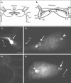
Octopamine-like immunoreactivity (OA-li) in the buccal ganglia of Aplysia and Helisoma. A1 Schematic representation of OA-li in the paired buccal ganglion of Aplysia. Results of experiments performed on two species, Aplysia californica and Aplysia dactylomela (shown here) were indistinguishable. Strong OA-li was limited to three moderately sized neurons near the caudal surface. A2 Two of the OA-li neurons were positioned symmetrically in the medial region of each hemiganglion (right hemiganglion shown here). These cells gave rise to fibers that traversed the buccal commissure connecting the two hemiganglia. A3 A single OA-li fiber in the cerebral-buccal connective (Cb c.). B1 Schematic representation of OA-li somata on the caudal surface of the Helisoma buccal ganglia. A tracing of the neuritic processes of a Lucifer yellow-stained neuron N3a was superimposed onto the upper OA-li soma. B2 Photograph of OA-li on the caudal surface of the right buccal ganglion of Helisoma trivolvis. ET is to upper right, Cb c. to lower right. Two N3A neuronal somata (arrow) are seen next to the commissure, and one unidentified OA-li soma (arrowhead) is seen in the lateral part of the ganglion. B3 In the left buccal ganglion one N3a (arrow) was stained near the buccal commissure and one unidentified lateral cell (arrowhead) was stained. The location of the unpaired N3a somata is variable and can be in either the left or right buccal ganglion. s n. = Salivary nerve; e n. = esophageal nerve; ET = esophageal trunk; bn1 = buccal nerve 1; bn2 = buccal nerve 2; bn3 = buccal nerve 3; Cb c. = cerebral-buccal connective; LBN = laterobuccal nerve; VBN = ventrobuccal nerve.
Fig. 5
Fig. 5
Schematic summary of proposed actions of OA-li and SCP-li neurons. This diagram is simplified to emphasize the roles of the neurons considered in this article. The polymorphic feeding motor systems of Helisoma and Aplysia can generate many variants of the feeding sequences depicted. Upper panel: during a bite without food contact, SCP-li RMs (B101 cluster neurons of Helisoma and RM cells of Aplysia) receive minimal exteroceptive stimulation. Under these conditions, OA interneuron firing (smaller font) does not evoke maximal hyperretraction in Helisoma and may not occur in Aplysia. In this sequence of actions, referred to as ‘biting’ in Aplysia [see Cropper et al., 2004], closure of the radula, which can be initiated by several pathways [see e.g. Plummer and Kirk, 1990; Evans and Cropper, 1998; Jing et al., 2004], begins near the peak of protraction and is primarily associated with the retraction phase. Lower panel: when a bite does encounter food or exteroceptive resistance, the SCP mechanoafferents are activated during the late phase of protraction. In Helisoma, they act in an ‘influential’ fashion on the OA-li interneurons (dashed curved arrow), increasing the intensity and duration of their firing (bold text, compare to upper panel). In Helisoma, this causes a stronger and more prolonged hyperretraction phase. We propose that the RM cells of Aplysia will similarly promote strong activation of the OA-li neurons and enhanced hyperretraction movements. The well characterized direct actions of the Aplysia RM neurons on radula closer motor neurons [Rosen et al., 2000a; Shetreat-Klein and Cropper, 2004] will contribute to coordinating radula closure with the prolongation of radula retraction.

Similar articles

Cited by

References

    1. Alexeeva V, Borovikov D, Miller MW, Rosen SC, Cropper EC. Effect of a serotonergic extrinsic modulatory neuron (MCC) on radula mechanoafferent function in Aplysia. J Neurophysiol. 1998;80:1609–1622. - PubMed
    1. Arbas EA, Meinertzhagen IA, Shaw SR. Evolution in nervous systems. Annu Rev Neurosci. 1991;14:9–38. - PubMed
    1. Arnett BC (1996) Feeding and regurgitation: two modes of operation of the buccal central pattern generator in Helisoma PhD thesis, Chicago, IL: University of Illinois at Chicago.
    1. Arshavsky YI, Deliagina TG, Meizerov ES, Orlovsky GN, Panchin YV. Control of feeding movements in the freshwater snail, Planorbis corneus. I. Rhythmic neurons of buccal ganglia. Exp Brain Res. 1988;70:310–322. - PubMed
    1. Audesirk T, Audesirk G. Behavior of gastropod mollusks. In: Willows AOD, editor. The Mollusca. Neurobiology and Behavior, Part 1. Vol 8. Orlando, FL: Academic Press; 1985. pp. 2–94.

Publication types