. 2009:5:337.
doi: 10.1038/msb.2009.93. Epub 2009 Dec 22.

A global view of protein expression in human cells, tissues, and organs

Affiliations

A global view of protein expression in human cells, tissues, and organs

Fredrik Pontén et al. Mol Syst Biol. 2009.

Abstract

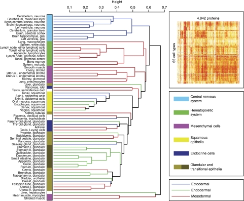
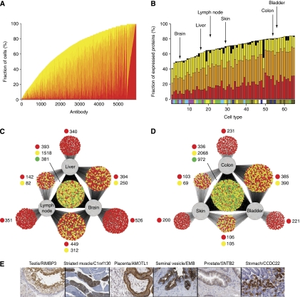
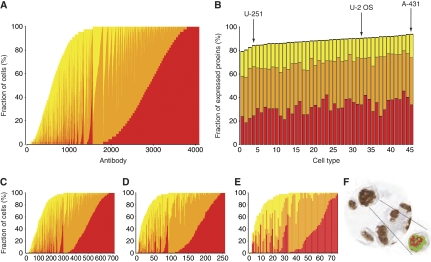
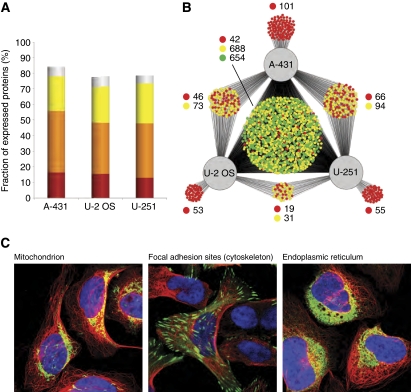
Defining the protein profiles of tissues and organs is critical to understanding the unique characteristics of the various cell types in the human body. In this study, we report on an anatomically comprehensive analysis of 4842 protein profiles in 48 human tissues and 45 human cell lines. A detailed analysis of over 2 million manually annotated, high-resolution, immunohistochemistry-based images showed a high fraction (>65%) of expressed proteins in most cells and tissues, with very few proteins (<2%) detected in any single cell type. Similarly, confocal microscopy in three human cell lines detected expression of more than 70% of the analyzed proteins. Despite this ubiquitous expression, hierarchical clustering analysis, based on global protein expression patterns, shows that the analyzed cells can be still subdivided into groups according to the current concepts of histology and cellular differentiation. This study suggests that tissue specificity is achieved by precise regulation of protein levels in space and time, and that different tissues in the body acquire their unique characteristics by controlling not which proteins are expressed but how much of each is produced.


Conflict of interest statement

The authors declare that they have no conflict of interest.

Figures

Figure 1
Figure 1
Global protein profiling in 65 normal human cell types. A dendrogram showing the relationships, based on global expression profiling, between the various cell types. The dendrogram was constructed using a hierarchical clustering model; the inset shows the original heat map. The underlying data are based on manual annotation of protein expression patterns in 65 normal cell types using 5934 antibodies corresponding to 4842 proteins. The dendrogram bars are labeled according to the proposed origin within the embryonic germ cell layers: ectoderm (blue), mesoderm (red), and endoderm (green). The cell types have been classified into six categories according to the color code to the right. A list of all the cell types can be found in Supplementary Table S2. The dendrograms for proteins encoded on single chromosomes are shown in Supplementary Figures S1–S3 and for random sets of 200 antibodies in Supplementary Figures S4–S6.
Figure 2
Figure 2
The tissue-specific protein expression in 65 cell types corresponding to 48 tissues and organs. (A) The fraction (%) of cells in which a particular protein was detected, including the fraction of cells with the relative expression levels strong (red), moderate (orange), and weak (yellow). A total of 5934 antibodies against 4842 proteins are arranged according to abundance of the corresponding protein target with cell-type-specific proteins to the left and ‘housekeeping proteins' to the right. The results for the various subfractions of antibodies are presented in Supplementary Figure S7. (B) The fraction (%) of the analyzed proteins detected in a specific cell type. Cells are arranged according to the fraction of proteins detected. A bar displaying the different color codes representing the six major categories of normal cell types (defined in Figure 1) is shown for each cell type. The name of each cell type is shown in Supplementary Table S2 and the results for the various subfractions of antibodies are presented in Supplementary Figure S8. Black represents missing data, i.e., where there was no representative cell type for a given immunostaining. The six cell types analyzed in C and D are pointed out by arrows. (C) A Cytoscape network plot (Shannon et al, 2003) showing the distribution of the analyzed proteins detected in at least one of the three cell types analyzed; liver hepatocytes, neurons of the cerebral cortex of the brain, and lymphoid cells from the germinal center of the lymph node. Each antibody/protein is represented by a small circle that is connected by a line/lines to the cells it was detected in. The color of the circle indicates the variability of staining intensity between the different cell lines; green indicates that all cell lines belonged to the same staining intensity category, yellow indicates that two cell lines belonged to the same staining intensity category, and red indicates that the staining intensity category was different for all three cell lines or only detected in a single cell. (D) A similar network plot based on the analysis of protein expression in glandular cells in the colon, epidermal cells from the skin, and urothelial cells from the bladder. (E) Six examples of proteins with essentially unknown functions that exhibit cell-type-specific expression. Testis—maturing spermatocytes and spermatids in the testicular seminiferous duct show strong partly membranous positivity with an antibody generated toward the uncharacterized protein RIMS-binding protein 3A. Muscle—striated skeletal muscle is shown with a fiber-type-specific sarcoplasmic positivity with an antibody directed toward an unknown protein encoded by C1orf130. Placenta—the expression of angiomotin-like protein 1 in placental tissue (immature) shows strong membranous positivity in basal cytotrophoblasts with moderate cytoplasmic positivity in syncytiotrophoblasts and exhibits distinct expression in the brush-border membrane. Seminal vesicle—glandular cells in the seminal vesicle were the only cells found to express embigin, a previously unknown protein. Prostate—in the prostate, moderate positivity was found with a membranous expression pattern for beta-2-syntrophin protein (SNTB2), a protein with unknown functions that has been shown to co-purify, with dystrophin, the protein product of the Duchenne muscular dystrophy locus. Stomach—in the stomach mucosa the previously unknown coiled-coil domain-containing protein 22 (CCDC22) was expressed in the parietal cells, producers of hydrochloric acid in response to histamine, acetylcholine, and gastrin.
Figure 3
Figure 3
Global protein expression in 45 human cell lines. (A) The fraction (%) of the 45 cell lines in which a particular protein was detected, including the fraction of the three relative expression levels: strong (red), moderate (orange), and weak (yellow). Each bar represents one of the 4096 antibodies with no missing data, i.e., where all cell lines were represented. (B) The fraction (%) of a total number of 5349 antibodies against 4349 proteins detected in a specific cell line, and with the cell lines ordered according to the fraction of proteins detected. The corresponding name and number of each cell line is shown in Supplementary Table S3 and the results for the various subfractions of antibodies are presented in Supplementary Figure S9. The same three staining categories were used and the black (top) part of the bar represents antibodies with missing data for the particular cell line. Arrows point out the three cell lines used in immunofluorescence analysis. (C) The fraction of cell lines in which each protein from a data set of 714 antibodies with supportive results from western blot analysis was detected. (D) A plot similar to C, with each bar representing one of the 257 antibodies remaining from a data set of paired HPA-antibodies, i.e., toward the same target protein, with no missing data for any of the cell lines, and a correlation coefficient of ⩾0.5 when cell line expression profiles were analyzed. (E) Same as D, but displaying only the results from the 75 antibodies with a correlation coefficient of ⩾0.8 and no missing data. (F) An example of cells, visualizing the interpretation of immunostaining by an automated image analysis software.
Figure 4
Figure 4
Global protein expression in three human cell lines using immunofluorescence-based confocal microscopy. (A) The fraction (%) of proteins detected in the three analyzed cell lines. The stainings are classified into the categories strong (red), moderate (orange), and weak (yellow) based on their measured intensity. Stainings annotated as weak with a granular cytoplasmic subcellular distribution (i.e., not a distinct cytoplasmic organelle or component) are considered less reliable (shown in gray). (B) A Cytoscape network plot (Shannon et al, 2003) showing the distribution of the analyzed proteins detected in at least one of the three cell lines. Each antibody/protein is represented by a small and the color of the circle indicates the variability of staining intensity between the different cell lines; green indicates that all cell lines belonged to the same staining intensity category, yellow indicates that two cell lines belonged to the same staining intensity category, and red indicates that the staining intensity category was different for all three cell lines. (C) Three example images of immunofluorescently stained U-2 OS showing proteins from the different categories (green, yellow, and red) in panel B and with different subcellular localizations. The protein of interest is shown in green, microtubules in red, and nuclei in blue. The first image (left) shows the 60-kDa heat shock protein (HSPD1) to be localized in the mitochondria and give a strong staining intensity in all three cell lines (green category) as detected by the antibody HPA001523. The second image (middle) shows the four and a half LIM domains protein 2 (FHL2) to be localized at focal adhesion sites in the cytoskeleton and give a strong staining intensity in U-2 OS and a moderate staining intensity in A-431 and U-251 MG (yellow category) as detected by the antibody HPA006028. The third image (right) shows the uncharacterized protein KIAA1467 to be localized in the endoplasmic reticulum and give a strong staining intensity in U-2 OS, moderate in A-431, and weak in U-251 MG (red category) as detected by the antibody HPA010803.

References

    1. Barbe L, Lundberg E, Oksvold P, Stenius A, Lewin E, Bjorling E, Asplund A, Ponten F, Brismar H, Uhlen M, Andersson-Svahn H (2008) Toward a confocal subcellular atlas of the human proteome. Mol Cell Proteomics 7: 499–508 - PubMed
    1. Berglund L, Bjorling E, Oksvold P, Fagerberg L, Asplund A, Szigyarto CA, Persson A, Ottosson J, Wernerus H, Nilsson P, Lundberg E, Sivertsson A, Navani S, Wester K, Kampf C, Hober S, Ponten F, Uhlen M (2008) A genecentric Human Protein Atlas for expression profiles based on antibodies. Mol Cell Proteomics 7: 2019–2027 - PubMed
    1. Birney E, Stamatoyannopoulos JA, Dutta A, Guigo R, Gingeras TR, Margulies EH, Weng Z, Snyder M, Dermitzakis ET, Thurman RE, Kuehn MS, Taylor CM, Neph S, Koch CM, Asthana S, Malhotra A, Adzhubei I, Greenbaum JA, Andrews RM, Flicek P et al. (2007) Identification and analysis of functional elements in 1% of the human genome by the ENCODE pilot project. Nature 447: 799–816 - PMC - PubMed
    1. Bjorling E, Lindskog C, Oksvold P, Linne J, Kampf C, Hober S, Uhlen M, Ponten F (2008) A web-based tool for in silico biomarker discovery based on tissue-specific protein profiles in normal and cancer tissues. Mol Cell Proteomics 7: 825–844 - PubMed
    1. Clamp M, Fry B, Kamal M, Xie X, Cuff J, Lin MF, Kellis M, Lindblad-Toh K, Lander ES (2007) Distinguishing protein-coding and noncoding genes in the human genome. Proc Natl Acad Sci USA 104: 19428–19433 - PMC - PubMed

Publication types

LinkOut - more resources