

Adoptive transfer of ex vivo HO-1 modified bone marrow-derived macrophages prevents liver ischemia and reperfusion injury
- PMID: 20029397
- PMCID: PMC2890105
- DOI: 10.1038/mt.2009.285
Adoptive transfer of ex vivo HO-1 modified bone marrow-derived macrophages prevents liver ischemia and reperfusion injury
Abstract
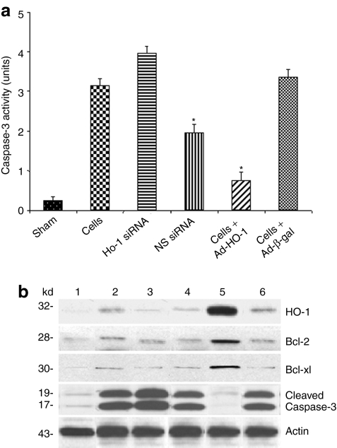
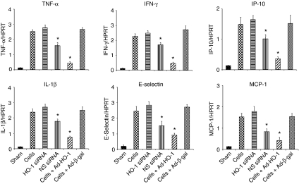
Macrophages play a critical role in the pathophysiology of liver ischemia and reperfusion (IR) injury (IRI). However, macrophages that overexpress antioxidant heme oxygenase-1 (HO-1) may exert profound anti-inflammatory functions. This study explores the cytoprotective effects and mechanisms of ex vivo modified HO-1-expressing bone marrow-derived macrophages (BMDMs) in well-defined mouse model of liver warm ischemia followed by reperfusion. Adoptive transfer of Ad-HO-1-transduced macrophages prevented IR-induced hepatocellular damage, as evidenced by depressed serum glutamic-oxaloacetic transaminase (sGOT) levels and preserved liver histology (Suzuki scores), compared to Ad-beta-gal controls. This beneficial effect was reversed following concomitant treatment with HO-1 siRNA. Ad-HO-1-transfected macrophages significantly decreased local neutrophil accumulation, TNF-alpha/IL-1beta, IFN-gamma/E-selectin, and IP-10/MCP-1 expression, caspase-3 activity, and the frequency of apoptotic cells, as compared with controls. Unlike in controls, Ad-HO-1-transfected macrophages markedly increased hepatic expression of antiapoptotic Bcl-2/Bcl-xl and depressed caspase-3 activity. These results establish the precedent for a novel investigative tool and provide the rationale for a clinically attractive new strategy in which native macrophages can be transfected ex vivo with cytoprotective HO-1 and then infused, if needed, to prospective recipients exposed to hepatic IR-mediated local inflammation, such as during liver transplantation, resection, or trauma.
Figures
Similar articles
-
Native macrophages genetically modified to express heme oxygenase 1 protect rat liver transplants from ischemia/reperfusion injury.Liver Transpl. 2011 Feb;17(2):201-10. doi: 10.1002/lt.22214. Liver Transpl. 2011. PMID: 21280193 Free PMC article.
-
Viral interleukin-10 gene transfer prevents liver ischemia-reperfusion injury: Toll-like receptor-4 and heme oxygenase-1 signaling in innate and adaptive immunity.Hum Gene Ther. 2007 Apr;18(4):355-66. doi: 10.1089/hum.2007.181. Hum Gene Ther. 2007. PMID: 17439357
-
Adoptive transfer of heme oxygenase-1 (HO-1)-modified macrophages rescues the nuclear factor erythroid 2-related factor (Nrf2) antiinflammatory phenotype in liver ischemia/reperfusion injury.Mol Med. 2014 Oct 14;20(1):448-55. doi: 10.2119/molmed.2014.00103. Mol Med. 2014. PMID: 25014792 Free PMC article.
-
Heme Oxygenase-1 in liver transplant ischemia-reperfusion injury: From bench-to-bedside.Free Radic Biol Med. 2020 Sep;157:75-82. doi: 10.1016/j.freeradbiomed.2020.02.012. Epub 2020 Feb 19. Free Radic Biol Med. 2020. PMID: 32084514 Free PMC article. Review.
-
Protective role of heme oxygenase-1 in fatty liver ischemia-reperfusion injury.Med Mol Morphol. 2019 Jun;52(2):61-72. doi: 10.1007/s00795-018-0205-z. Epub 2018 Aug 31. Med Mol Morphol. 2019. PMID: 30171344 Free PMC article. Review.
Cited by
-
Heme mediated STAT3 activation in severe malaria.PLoS One. 2012;7(3):e34280. doi: 10.1371/journal.pone.0034280. Epub 2012 Mar 30. PLoS One. 2012. PMID: 22479586 Free PMC article.
-
β-catenin regulates innate and adaptive immunity in mouse liver ischemia-reperfusion injury.Hepatology. 2013 Mar;57(3):1203-14. doi: 10.1002/hep.26100. Epub 2013 Feb 7. Hepatology. 2013. PMID: 23081841 Free PMC article.
-
Heme oxygenase system in hepatic ischemia-reperfusion injury.World J Gastroenterol. 2010 Dec 28;16(48):6068-78. doi: 10.3748/wjg.v16.i48.6068. World J Gastroenterol. 2010. PMID: 21182221 Free PMC article.
-
Heme oxygenase-1 regulates sirtuin-1-autophagy pathway in liver transplantation: From mouse to human.Am J Transplant. 2018 May;18(5):1110-1121. doi: 10.1111/ajt.14586. Epub 2017 Dec 18. Am J Transplant. 2018. PMID: 29136322 Free PMC article.
-
Dual Effect of Hepatic Macrophages on Liver Ischemia and Reperfusion Injury during Liver Transplantation.Immune Netw. 2018 Jun 28;18(3):e24. doi: 10.4110/in.2018.18.e24. eCollection 2018 Jun. Immune Netw. 2018. PMID: 29984042 Free PMC article. Review.
References
-
- Farmer DG, Amersi F, Kupiec-Weglinski JW., and , Busuttil RW. Current status of ischemia and reperfusion injury in the liver. Transplant Rev. 2000;14:106–126.
-
- Teoh NC., and , Farrell GC. Hepatic ischemia reperfusion injury: pathogenic mechanisms and basis for hepatoprotection. J Gastroenterol Hepatol. 2003;18:891–902. - PubMed
-
- Ozaki M, Suzuki S., and , Irani K. Redox factor-1/APE suppresses oxidative stress by inhibiting the rac1 GTPase. FASEB J. 2002;16:889–890. - PubMed
-
- Li C., and , Jackson RM. Reactive species mechanisms of cellular hypoxia-reoxygenation injury. Am J Physiol Cell Physiol. 2002;282:C227–C241. - PubMed
Publication types
MeSH terms
Substances
Grants and funding
LinkOut - more resources
Full Text Sources
Research Materials
Miscellaneous
