

Early nonischemic oxidative metabolic dysfunction leads to chronic brain atrophy in traumatic brain injury
- PMID: 20029449
- PMCID: PMC2949156
- DOI: 10.1038/jcbfm.2009.263
Early nonischemic oxidative metabolic dysfunction leads to chronic brain atrophy in traumatic brain injury
Abstract
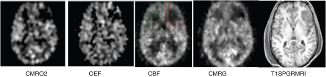
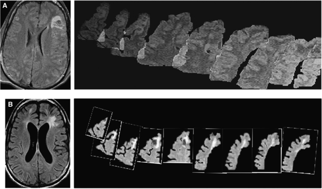
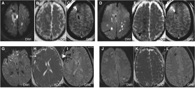
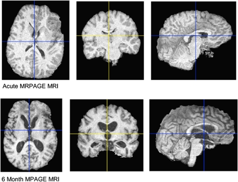
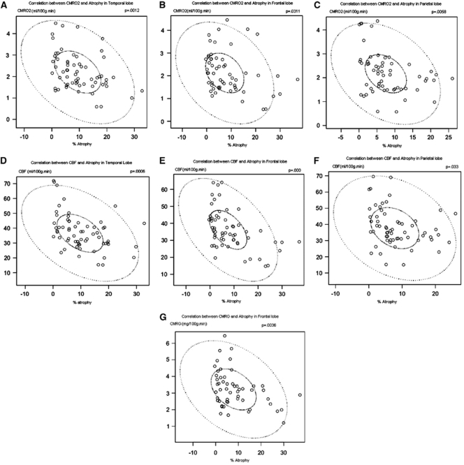
Chronic brain atrophy after traumatic brain injury (TBI) is a well-known phenomenon, the causes of which are unknown. Early nonischemic reduction in oxidative metabolism is regionally associated with chronic brain atrophy after TBI. A total of 32 patients with moderate-to-severe TBI prospectively underwent positron emission tomography (PET) and volumetric magnetic resonance imaging (MRI) within the first week and at 6 months after injury. Regional lobar assessments comprised oxidative metabolism and glucose metabolism. Acute MRI showed a preponderance of hemorrhagic lesions with few irreversible ischemic lesions. Global and regional chronic brain atrophy occurred in all patients by 6 months, with the temporal and frontal lobes exhibiting the most atrophy compared with the occipital lobe. Global and regional reduction in cerebral metabolic rate of oxygen (CMRO(2)), cerebral blood flow (CBF), oxygen extraction fraction (OEF), and cerebral metabolic rate of glucose were observed. The extent of metabolic dysfunction was correlated with the total hemorrhage burden on initial MRI (r=0.62, P=0.01). The extent of regional brain atrophy correlated best with CMRO(2) and CBF. Lobar values of OEF were not in the ischemic range and did not correlate with chronic brain atrophy. Chronic brain atrophy is regionally specific and associated with regional reductions in oxidative brain metabolism in the absence of irreversible ischemia.
Figures
Similar articles
-
Regional cerebrovascular and metabolic effects of hyperventilation after severe traumatic brain injury.J Neurosurg. 2002 Jan;96(1):103-8. doi: 10.3171/jns.2002.96.1.0103. J Neurosurg. 2002. PMID: 11794590
-
Physiological thresholds for irreversible tissue damage in contusional regions following traumatic brain injury.Brain. 2005 Aug;128(Pt 8):1931-42. doi: 10.1093/brain/awh536. Epub 2005 May 11. Brain. 2005. PMID: 15888537
-
Early metabolic characteristics of lesion and nonlesion tissue after head injury.J Cereb Blood Flow Metab. 2009 May;29(5):965-75. doi: 10.1038/jcbfm.2009.22. Epub 2009 Mar 18. J Cereb Blood Flow Metab. 2009. PMID: 19293825
-
[Cerebral oxygen consumption and ischemia in traumatic brain injury].Minerva Anestesiol. 2004 Apr;70(4):207-11. Minerva Anestesiol. 2004. PMID: 15173697 Review. Italian.
-
Capillary transit time heterogeneity and flow-metabolism coupling after traumatic brain injury.J Cereb Blood Flow Metab. 2014 Oct;34(10):1585-98. doi: 10.1038/jcbfm.2014.131. Epub 2014 Jul 23. J Cereb Blood Flow Metab. 2014. PMID: 25052556 Free PMC article. Review.
Cited by
-
Alterations in cerebral oxygen metabolism after traumatic brain injury in children.J Cereb Blood Flow Metab. 2013 Jan;33(1):48-52. doi: 10.1038/jcbfm.2012.130. Epub 2012 Sep 12. J Cereb Blood Flow Metab. 2013. PMID: 22968318 Free PMC article.
-
High blood glucose does not adversely affect outcome in moderately brain-injured rodents.J Neurotrauma. 2010 Aug;27(8):1439-48. doi: 10.1089/neu.2010.1328. J Neurotrauma. 2010. PMID: 20504157 Free PMC article.
-
Transplantation of marrow stromal cells restores cerebral blood flow and reduces cerebral atrophy in rats with traumatic brain injury: in vivo MRI study.J Neurotrauma. 2011 Apr;28(4):535-45. doi: 10.1089/neu.2010.1619. Epub 2011 Mar 24. J Neurotrauma. 2011. PMID: 21275806 Free PMC article.
-
Deferoxamine attenuates acute hydrocephalus after traumatic brain injury in rats.Transl Stroke Res. 2014 Oct;5(5):586-94. doi: 10.1007/s12975-014-0353-y. Epub 2014 Jun 17. Transl Stroke Res. 2014. PMID: 24935175 Free PMC article.
-
Theory of mind mediates the prospective relationship between abnormal social brain network morphology and chronic behavior problems after pediatric traumatic brain injury.Soc Cogn Affect Neurosci. 2016 Apr;11(4):683-92. doi: 10.1093/scan/nsw007. Epub 2016 Jan 21. Soc Cogn Affect Neurosci. 2016. PMID: 26796967 Free PMC article.
References
-
- Bergsneider M, Hovda DA, McArthur DL, Etchepare M, Huang SC, Sehati N, Satz P, Phelps ME, Becker DP. Metabolic recovery following human traumatic brain injury based on FDG-PET: time course and relationship to neurological disability. J Head Trauma Rehabil. 2001;16:135–148. - PubMed
-
- Coles JP, Cunningham AS, Salvador R, Chatfield DA, Carpenter A, Pickard JD, Menon DK. Early metabolic characteristics of lesion and nonlesion tissue after head injury. J Cereb Blood Flow Metab. 2009;29:965–975. - PubMed
-
- Coles JP, Fryer TD, Smielewski P, Rice K, Clark JC, Pickard JD, Menon DK. Defining ischemic burden after traumatic brain injury using 15O PET imaging of cerebral physiology. J Cereb Blood Flow Metab. 2004;24:191–201. - PubMed
-
- Cunningham AS, Salvador R, Coles JP, Chatfield DA, Bradley PG, Johnston AJ, Steiner LA, Fryer TD, Aigbirhio FI, Smielewski P, Williams GB, Carpenter TA, Gillard JH, Pickard JD, Menon AK. Physiological thresholds for irreversible tissue damage in contusional regions following traumatic brain injury. Brain. 2005;128:1931–1942. - PubMed
-
- Dinov ID, Van Horn JD, Lozev KM, Magsipoc R, Petrosyan P, Liu Z, MacKenzie-Graham A, Eggert P, Parker DS, Toga AW.2009Efficient, distributed and interactive neuroimaging data analysis using the LONI pipelineFront. Neuroinform. 3:22. doi:10.3389/neuro.11.022.2009published online: 20 July 2009 - DOI - PMC - PubMed
Publication types
MeSH terms
Substances
Grants and funding
LinkOut - more resources
Full Text Sources
Medical
