

Agmatine attenuates brain edema through reducing the expression of aquaporin-1 after cerebral ischemia
- PMID: 20029450
- PMCID: PMC2949179
- DOI: 10.1038/jcbfm.2009.260
Agmatine attenuates brain edema through reducing the expression of aquaporin-1 after cerebral ischemia
Abstract
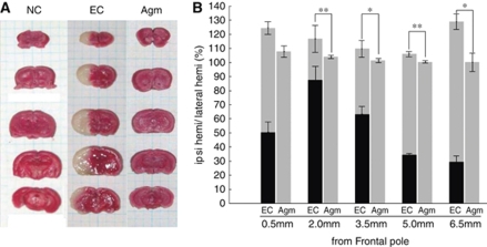
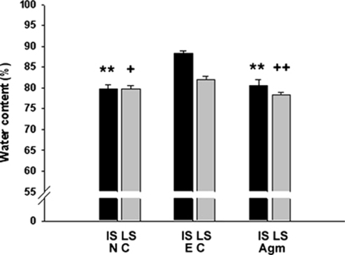
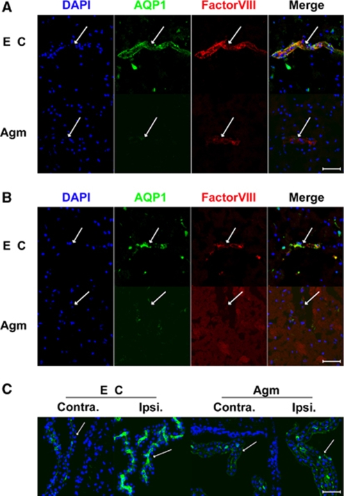
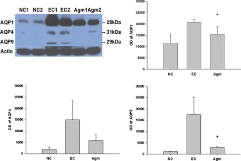
Brain edema is frequently shown after cerebral ischemia. It is an expansion of brain volume because of increasing water content in brain. It causes to increase mortality after stroke. Agmatine, formed by the decarboxylation of L-arginine by arginine decarboxylase, has been shown to be neuroprotective in trauma and ischemia models. The purpose of this study was to investigate the effect of agmatine for brain edema in ischemic brain damage and to evaluate the expression of aquaporins (AQPs). Results showed that agmatine significantly reduced brain swelling volume 22 h after 2 h middle cerebral artery occlusion in mice. Water content in brain tissue was clearly decreased 24 h after ischemic injury by agmatine treatment. Blood-brain barrier (BBB) disruption was diminished with agmatine than without. The expressions of AQPs-1 and -9 were well correlated with brain edema as water channels, were significantly decreased by agmatine treatment. It can thus be suggested that agmatine could attenuate brain edema by limiting BBB disruption and blocking the accumulation of brain water content through lessening the expression of AQP-1 after cerebral ischemia.
Figures
Similar articles
-
Effect of propofol post-treatment on blood-brain barrier integrity and cerebral edema after transient cerebral ischemia in rats.Neurochem Res. 2013 Nov;38(11):2276-86. doi: 10.1007/s11064-013-1136-7. Epub 2013 Aug 29. Neurochem Res. 2013. PMID: 23990224
-
Neuroprotective effect of agmatine in rats with transient cerebral ischemia using MR imaging and histopathologic evaluation.Magn Reson Imaging. 2013 Sep;31(7):1174-81. doi: 10.1016/j.mri.2013.03.026. Epub 2013 May 1. Magn Reson Imaging. 2013. PMID: 23642800
-
The effect of ASK1 on vascular permeability and edema formation in cerebral ischemia.Brain Res. 2015 Jan 21;1595:143-55. doi: 10.1016/j.brainres.2014.11.024. Epub 2014 Nov 15. Brain Res. 2015. PMID: 25446452
-
Role of agmatine in neurodegenerative diseases and epilepsy.Front Biosci (Elite Ed). 2014 Jun 1;6(2):341-59. doi: 10.2741/E710. Front Biosci (Elite Ed). 2014. PMID: 24896210 Review.
-
Treatment Effects of Acetazolamide on Ischemic Stroke: A Meta-Analysis and Systematic Review.World Neurosurg. 2024 May;185:e750-e757. doi: 10.1016/j.wneu.2024.02.123. Epub 2024 Feb 28. World Neurosurg. 2024. PMID: 38423457
Cited by
-
Agmatine Modulates the Phenotype of Macrophage Acute Phase after Spinal Cord Injury in Rats.Exp Neurobiol. 2017 Oct;26(5):278-286. doi: 10.5607/en.2017.26.5.278. Epub 2017 Oct 16. Exp Neurobiol. 2017. PMID: 29093636 Free PMC article.
-
Neural Stem Cells Overexpressing Arginine Decarboxylase Improve Functional Recovery from Spinal Cord Injury in a Mouse Model.Int J Mol Sci. 2022 Dec 13;23(24):15784. doi: 10.3390/ijms232415784. Int J Mol Sci. 2022. PMID: 36555425 Free PMC article.
-
Agmatine Attenuates Brain Edema and Apoptotic Cell Death after Traumatic Brain Injury.J Korean Med Sci. 2015 Jul;30(7):943-52. doi: 10.3346/jkms.2015.30.7.943. Epub 2015 Jun 10. J Korean Med Sci. 2015. PMID: 26130959 Free PMC article.
-
Retroviral expression of human arginine decarboxylase reduces oxidative stress injury in mouse cortical astrocytes.BMC Neurosci. 2014 Aug 26;15:99. doi: 10.1186/1471-2202-15-99. BMC Neurosci. 2014. PMID: 25156824 Free PMC article.
-
M2 Phenotype Microglia-derived Cytokine Stimulates Proliferation and Neuronal Differentiation of Endogenous Stem Cells in Ischemic Brain.Exp Neurobiol. 2017 Feb;26(1):33-41. doi: 10.5607/en.2017.26.1.33. Epub 2017 Feb 3. Exp Neurobiol. 2017. PMID: 28243165 Free PMC article.
References
-
- Agre P, Nielsen S, Ottersen OP. Towards a molecular understanding of water homeostasis in the brain. Neuroscience. 2004;129:849–850. - PubMed
-
- Amiry-Moghaddam M, Ottersen OP. The molecular basis of water transport in the brain. Nat Rev Neurosci. 2003;4:991–1001. - PubMed
-
- Auguet M, Viossat I, Marin JG, Chabrier PE. Selective inhibition of inducible nitric oxide synthase by agmatine. Jpn J Pharmacol. 1995;69:285–287. - PubMed
-
- Badaut J, Hirt L, Granziera C, Bogousslavsky J, Magistretti PJ, Regli L. Astrocyte-specific expression of aquaporin-9 in mouse brain is increased after transient focal cerebral ischemia. J Cereb Blood Flow Metab. 2001;21:477–482. - PubMed
Publication types
MeSH terms
Substances
LinkOut - more resources
Full Text Sources
