

Hypoxia induces T-cell apoptosis by inhibiting chemokine C receptor 7 expression: the role of adenosine receptor A(2)
- PMID: 20029460
- PMCID: PMC4003256
- DOI: 10.1038/cmi.2009.105
Hypoxia induces T-cell apoptosis by inhibiting chemokine C receptor 7 expression: the role of adenosine receptor A(2)
Abstract
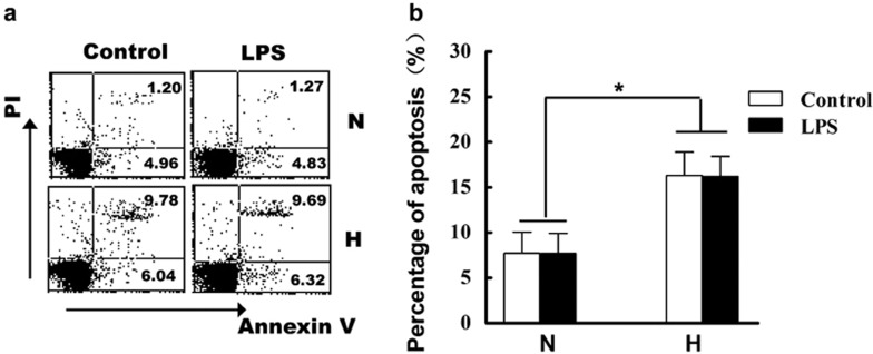
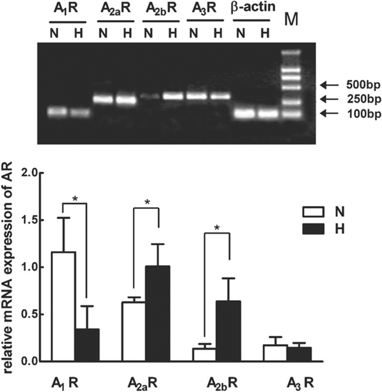
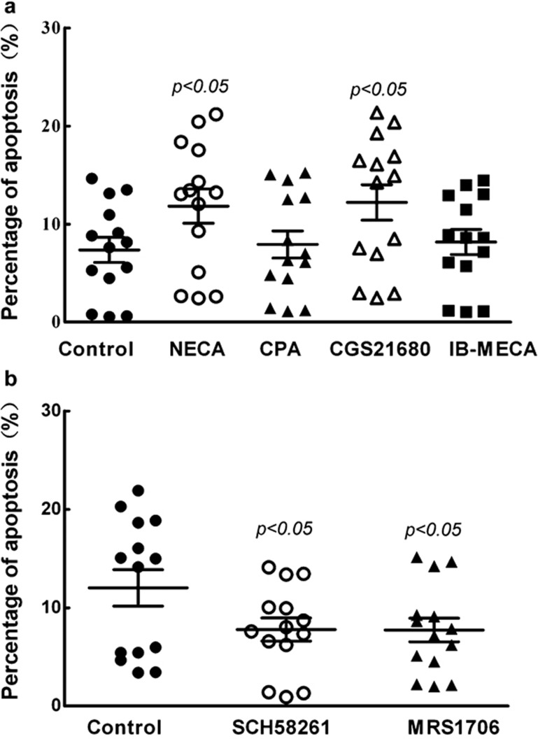
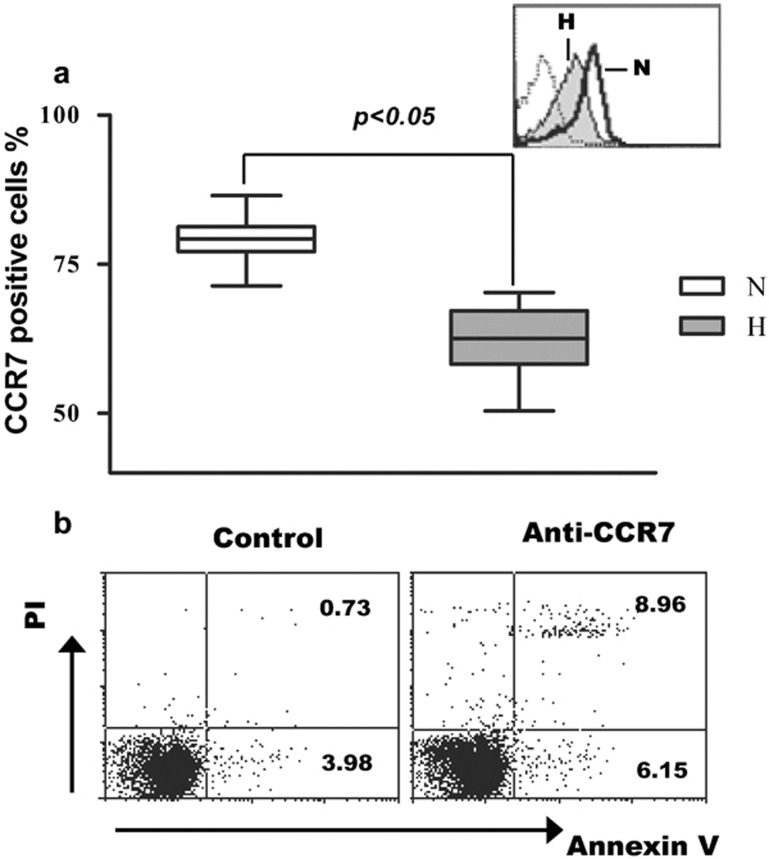
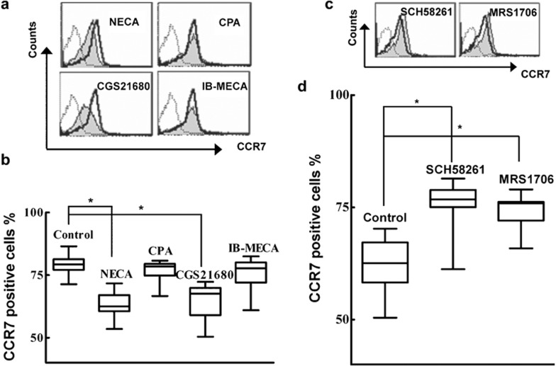
Hypoxia is a major characteristic of the tumor microenvironment, and its effects on immune cells are proposed to be important factors for the process of tumor immune escape. It has been reported that hypoxia affects the function of dendritic cells and the antitumor function of T cells. Here we discuss the effects of hypoxia on T-cell survival. Our results showed that hypoxia induced apoptosis of T cells. Adenosine and adenosine receptors (AR) are important to the hypoxia-related signaling pathway. Using AR agonists and antagonists, we demonstrated that hypoxia-induced apoptosis of T cells was mediated by A(2a )and A(2b) receptors. Furthermore, we are the first, to our knowledge, to report that hypoxia significantly inhibited the expression of chemokine C receptor 7 (CCR7) of T cells via the A(2)R signal pathway, perhaps representing a mechanism of hypoxia-induced apoptosis of T cells. Collectively, our research demonstrated that hypoxia induces T-cell apoptosis by the A(2)R signaling pathway partly by suppressing CCR7. Blocking the A(2)R signaling pathway and/or activation of CCR7 can increase the anti-apoptosis function of T cells and may become a new strategy to improve antitumor potential.
Figures
Similar articles
-
HIF-dependent induction of adenosine receptor A2b skews human dendritic cells to a Th2-stimulating phenotype under hypoxia.Immunol Cell Biol. 2010 Feb;88(2):165-71. doi: 10.1038/icb.2009.77. Epub 2009 Oct 20. Immunol Cell Biol. 2010. PMID: 19841638
-
NF-κB enhances hypoxia-driven T-cell immunosuppression via upregulation of adenosine A(2A) receptors.Cell Signal. 2014 May;26(5):1060-7. doi: 10.1016/j.cellsig.2014.01.024. Epub 2014 Jan 29. Cell Signal. 2014. PMID: 24486403
-
T regulatory cells: hypoxia-adenosinergic suppression and re-direction of the immune response.Trends Immunol. 2009 Mar;30(3):102-8. doi: 10.1016/j.it.2008.12.002. Epub 2009 Feb 7. Trends Immunol. 2009. PMID: 19201652
-
Targeting Hypoxia-A2A Adenosinergic Immunosuppression of Antitumor T Cells During Cancer Immunotherapy.Front Immunol. 2020 Sep 29;11:570041. doi: 10.3389/fimmu.2020.570041. eCollection 2020. Front Immunol. 2020. PMID: 33117358 Free PMC article. Review.
-
Regulation of lymphocyte function by adenosine.Arterioscler Thromb Vasc Biol. 2012 Sep;32(9):2097-103. doi: 10.1161/ATVBAHA.111.226837. Epub 2012 Jul 5. Arterioscler Thromb Vasc Biol. 2012. PMID: 22772752 Free PMC article. Review.
Cited by
-
Adenosine Induces EBV Lytic Reactivation through ADORA1 in EBV-Associated Gastric Carcinoma.Int J Mol Sci. 2019 Mar 14;20(6):1286. doi: 10.3390/ijms20061286. Int J Mol Sci. 2019. PMID: 30875759 Free PMC article.
-
Mechanisms regulating immune surveillance of cellular stress in cancer.Cell Mol Life Sci. 2018 Jan;75(2):225-240. doi: 10.1007/s00018-017-2597-7. Epub 2017 Jul 25. Cell Mol Life Sci. 2018. PMID: 28744671 Free PMC article. Review.
-
The Therapeutic Potential of Physical Exercise in Cancer: The Role of Chemokines.Int J Mol Sci. 2024 Dec 23;25(24):13740. doi: 10.3390/ijms252413740. Int J Mol Sci. 2024. PMID: 39769501 Free PMC article. Review.
-
Age-Related Gene Alteration in Naïve and Memory T cells Using Precise Age-Tracking Model.Front Cell Dev Biol. 2021 Feb 11;8:624380. doi: 10.3389/fcell.2020.624380. eCollection 2020. Front Cell Dev Biol. 2021. PMID: 33644036 Free PMC article.
-
Cancer Cell Metabolism Bolsters Immunotherapy Resistance by Promoting an Immunosuppressive Tumor Microenvironment.Front Oncol. 2020 Jul 22;10:1197. doi: 10.3389/fonc.2020.01197. eCollection 2020. Front Oncol. 2020. PMID: 32775303 Free PMC article. Review.
References
-
- Hoskin DW, Mader JS, Furlong SJ, Conrad DM, Blay J. Inhibition of T cell and natural killer cell function by adenosine and its contribution to immune evasion by tumor cells (Review) Int J Oncol. 2008;32:527–535. - PubMed
-
- Sitkovsky M, Lukashev D. Regulation of immune cells by local-tissue oxygen tension: HIF1 alpha and adenosine receptors. Nat Rev Immunol. 2005;5:712–721. - PubMed
-
- Helmlinger G, Yuan F, Dellian M, Jain RK. Interstitial pH and pO2 gradients in solid tumors in vivo: high-resolution measurements reveal a lack of correlation. Nat Med. 1997;3:177–182. - PubMed
-
- Zhao P, Li XG, Yang M, Shao Q, Wang D, Liu S, et al. Hypoxia suppresses the production of MMP-9 by human monocyte-derived dendritic cells and requires activation of adenosine receptor A2b via cAMP/PKA signaling pathway. Mol Immunol. 2008;45:2187–2195. - PubMed
-
- Qu X, Yang MX, Kong BH, Qi L, Lam QL, Yan S, et al. Hypoxia inhibits the migratory capacity of human monocyte-derived dendritic cells. Immunol Cell Biol. 2005;83:668–673. - PubMed
Publication types
MeSH terms
Substances
LinkOut - more resources
Full Text Sources
Research Materials
