Review
. 2009:2009:135249.
doi: 10.1155/2009/135249. Epub 2009 Dec 8.

Role of ryanodine receptor subtypes in initiation and formation of calcium sparks in arterial smooth muscle: comparison with striated muscle

Affiliations
Review

Role of ryanodine receptor subtypes in initiation and formation of calcium sparks in arterial smooth muscle: comparison with striated muscle

Kirill Essin et al. J Biomed Biotechnol. 2009.

Abstract

Calcium sparks represent local, rapid, and transient calcium release events from a cluster of ryanodine receptors (RyRs) in the sarcoplasmic reticulum. In arterial smooth muscle cells (SMCs), calcium sparks activate calcium-dependent potassium channels causing decrease in the global intracellular [Ca2+] and oppose vasoconstriction. This is in contrast to cardiac and skeletal muscle, where spatial and temporal summation of calcium sparks leads to global increases in intracellular [Ca2+] and myocyte contraction. We summarize the present data on local RyR calcium signaling in arterial SMCs in comparison to striated muscle and muscle-specific differences in coupling between L-type calcium channels and RyRs. Accordingly, arterial SMC Ca(v)1.2 L-type channels regulate intracellular calcium stores content, which in turn modulates calcium efflux though RyRs. Downregulation of RyR2 up to a certain degree is compensated by increased SR calcium content to normalize calcium sparks. This indirect coupling between Ca(v)1.2 and RyR in arterial SMCs is opposite to striated muscle, where triggering of calcium sparks is controlled by rapid and direct cross-talk between Ca(v)1.1/Ca(v)1.2 L-type channels and RyRs. We discuss the role of RyR isoforms in initiation and formation of calcium sparks in SMCs and their possible molecular binding partners and regulators, which differ compared to striated muscle.


Figures
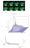
Figure 1
Figure 1
Calcium spark in a rat tibial artery smooth muscle cell. (a) Series of 8 consecutive laser scanning confocal images (17 milliseconds apart) illustrating a calcium spark. The calcium spark area is marked by a square and the peak of the calcium spark is indicated by arrows (b). The myocyte was loaded with the Ca2+ indicator dye fluo-3. Two-dimensional (2D) images were obtained using a Nipkow spinning disk confocal microscope (Perkin Elmer, UltraView LCI). (b) Three-dimensional plot of fluorescence intensity of the cell shown in (a) at 68 milliseconds. The calcium spark occurred in close proximity to the plasma membrane. (c) Time course of the calcium spark in the marked area. The amplitude is expressed as F/Fo. F is the fluorescence intensity in the marked area where the spark appeared. Fo is fluorescence intensity of the same cell area in the absence of calcium spark.
Figure 2
Figure 2
Control of calcium sparks by voltage-dependent L-type calcium channels. (a) Ca2+ entry from extracellular space is not required for calcium release in skeletal muscle. Charge movement within Cav1.1 activates RyR1 channel via a direct physical interaction. Ca2+ efflux from SR through the opened RyR1 channel activates nearby RyR (RyR3) channels via calcium-induced calcium release (CICR). (b) In cardiomyocytes Cav1.2 mediates influx of extracellular Ca2+ into cytosol. Ca2+ then binds to and activates RyR2 channels via CICR. (c) Triggering of calcium sparks is not controlled by rapid, direct cross-talk between Cav1.2 channels and RyRs in smooth muscle cells in contrast to cardiac and skeletal muscle cells. Instead, Cav1.2 channels contribute to global cytosolic [Ca2+], which in turn influences luminal SR calcium and thus calcium sparks. SERCA: sarco/endoplasmic reticulum calcium ATPase, BK: big conductance calcium-activated K+ channel.
Figure 3
Figure 3
In vivo suppression of RyR2 expression by antisense oligonucleotides (AS ODNs) in rat cerebral artery smooth muscle cells leads to increase of SR Ca2+ content without effects on spontaneous calcium sparks. (a) Left side: Images of rat cerebral arteries isolated 7 days after continuous i.c.v. injection of the fluorescence-labled AS ODNs (left) and nonlabeled AS ODNs (right) via Alzet osmotic pumps. Right side: RyR2 mRNA expression normalized to 18S ribosomal mRNA expression in arteries isolated from rats treated by AS ODNs (red) and scrambled (SC) ODNs (black) (n = 5 rats in each group). (b) Left side: Averaged time course of changes in response to external 10 mM caffeine in fluorescence intensity in Fluo-4 loaded cerebral artery smooth muscle cells (SMCs) isolated from rats treated with AS ODNs (red) and SC ODNs (black). The corresponding mean peak amplitudes are presented in the middle plot. Right side: The frequency of calcium sparks was not different between AS and SC ODNs treated cells. Calcium sparks were recorded in line-scan mode. The number of cells tested is indicated above each bar. Data are mean ± S.E.M. AU: arbitrary unit.
Figure 4
Figure 4
Putative role of sorcin, homer1, and calstabins in calcium spark regulation in smooth muscle. Sorcin (soluble resistance-related Ca2+ binding protein) is a negative regulator of calcium sparks in SMCs. It is activated by increases in cytosolic [Ca2+] (EC50  = 1.5 μM). Stimulatory effects on Cav1.2 channels and SERCA have also been reported. Calstabin2 (FKBP12.6) associates with RyR2 and stabilizes RyR2 channels in their closed state. FK506, an immunodepressant drug, binds to calstabin2 and can cause dissociation of calstabin2-RyR2 complex and therefore stimulation of calcium sparks in some smooth muscle tissues. Scaffold protein Homer1 (Ves1) has been shown to stimulate both RyR2 (at doses <50 nM) and Cav1.2 channels in nonarterial smooth muscle. Based on these findings, Homer1 can be expected to be a positive regulator of local calcium release calcium in this type of smooth muscle, which, however, has not been demonstrated so far. With respect to arterial smooth muscle, it is likely that calstabin2 and Homer1 play no role or only a minor role in calcium spark generation. This speculation is based on our findings that cytosolic [Ca2+] itself contributes minimally to the acute triggering of physiologically relevant proportion of calcium sparks. Instead the most efficacious calcium spark trigger appears to be the luminal SR Ca2+, which is slowly loaded via Ca2+ influx through Cav1.2 channels. Arrows indicate positive (red, +) and negative (blue, −) regulation of corresponding targets.

Similar articles

Cited by

References

    1. Cheng H, Lederer WJ. Calcium sparks. Physiological Reviews. 2008;88(4):1491–1545. - PubMed
    1. Benham CD, Bolton TB. Spontaneous transient outward currents in single visceral and vascular smooth muscle cells of the rabbit. Journal of Physiology. 1986;381:385–406. - PMC - PubMed
    1. Nelson MT, Cheng H, Rubart M, et al. Relaxation of arterial smooth muscle by calcium sparks. Science. 1995;270(5236):633–637. - PubMed
    1. Perez GJ, Bonev AD, Patlak JB, Nelson MT. Functional coupling of ryanodine receptors to K(Ca) channels in smooth muscle cells from rat cerebral arteries. Journal of General Physiology. 1999;113(2):229–237. - PMC - PubMed
    1. Ledoux J, Werner ME, Brayden JE, Nelson MT. Calcium-activated potassium channels and the regulation of vascular tone. Physiology. 2006;21(1):69–79. - PubMed

Publication types

MeSH terms

Substances