

Atypical craniosynostosis with torticollis and neurological symptoms: a rhombencephalosynapsis sequence
- PMID: 20029674
- PMCID: PMC2796237
- DOI: 10.1155/2009/919463
Atypical craniosynostosis with torticollis and neurological symptoms: a rhombencephalosynapsis sequence
Abstract
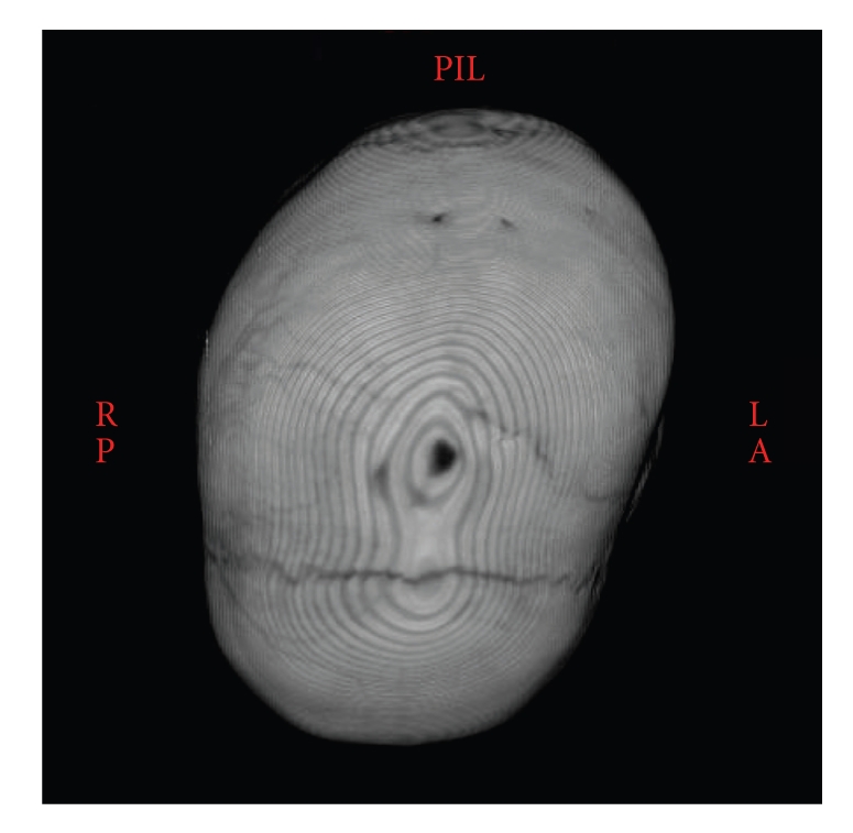
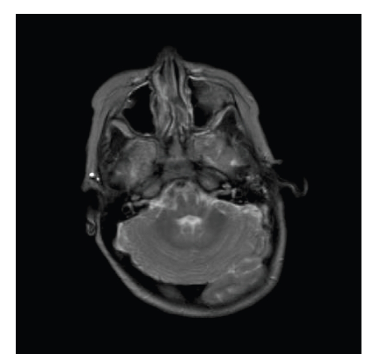
Purpose. We describe a case of 3-year-old girl with rhombencephalosynapsis, a rare cerebellar anomaly. Patient. A 3-year-old girl was admitted to our hospital due to congenital torticollis and asymmetry of face, skull and trunk. Craniosynostosis was suspected due to abnormal head shape. 3D-CT revealed closure of the sagittal suture without scaphocephalic skull. Due to atypical craniosynostosis with neurological symptoms, brain-MRI was performed revealing rhombencephalosynapsis. Results. Our patient presented with atypical craniosynostosis and balance problems, not typical for scaphocephaly. Operative treatment for craniosynotosis was not carried out because the cause of the problems was the cerebellum instead of the brain. Conclusions. Therefore, we conclude that patients with atypical craniosynostosis should be examined with brain-MRI to exclude the intracranial malformations, which 3D-CT does not reveal. Without brain-MRI, decision (not) to perform surgery could have been different.
Figures
Similar articles
-
Congenital muscular torticollis concurrent with sagittal synostosis: a case report.Ann Rehabil Med. 2014 Oct;38(5):712-6. doi: 10.5535/arm.2014.38.5.712. Epub 2014 Oct 30. Ann Rehabil Med. 2014. PMID: 25379504 Free PMC article.
-
A Treatment Algorithm for Patients Presenting with Sagittal Craniosynostosis after the Age of 1 Year.Plast Reconstr Surg. 2017 Sep;140(3):582-590. doi: 10.1097/PRS.0000000000003602. Plast Reconstr Surg. 2017. PMID: 28841620
-
Isolated Sagittal Craniosynostosis: A Comprehensive Review.Diagnostics (Basel). 2024 Feb 16;14(4):435. doi: 10.3390/diagnostics14040435. Diagnostics (Basel). 2024. PMID: 38396475 Free PMC article. Review.
-
Comparison of Black Bone MRI and 3D-CT in the preoperative evaluation of patients with craniosynostosis.J Plast Reconstr Aesthet Surg. 2020 Apr;73(4):723-731. doi: 10.1016/j.bjps.2019.11.006. Epub 2019 Nov 27. J Plast Reconstr Aesthet Surg. 2020. PMID: 31917189
-
Craniosynostosis: prenatal diagnosis by means of ultrasound and SSSE-MRI. Family series with report of neurodevelopmental outcome and review of the literature.Arch Gynecol Obstet. 2011 Apr;283(4):909-16. doi: 10.1007/s00404-010-1643-6. Epub 2010 Sep 2. Arch Gynecol Obstet. 2011. PMID: 20811900 Review.
Cited by
-
Congenital muscular torticollis concurrent with sagittal synostosis: a case report.Ann Rehabil Med. 2014 Oct;38(5):712-6. doi: 10.5535/arm.2014.38.5.712. Epub 2014 Oct 30. Ann Rehabil Med. 2014. PMID: 25379504 Free PMC article.
-
Rhombencephalosynapsis With Obstructive Hydrocephalus: A Rare Presentation of the Cerebellar Anomaly on MRI Findings.Cureus. 2023 Jun 5;15(6):e39969. doi: 10.7759/cureus.39969. eCollection 2023 Jun. Cureus. 2023. PMID: 37416012 Free PMC article.
References
-
- Simmons G, Damiano TR, Truwit CL. MRI and clinical findings in rhombencephalosynapsis. Journal of Computer Assisted Tomography. 1993;17(2):211–214. - PubMed
-
- Danon O, Elmaleh M, Boukobza B, Fohlen M, Hadjnacer K, Hassan M. Rhombencephalosynapsis diagnosed in childhood: clinical and MRI findings. Magnetic Resonance Imaging. 2000;18(1):99–101. - PubMed
-
- Obersteiner H. Ein kleinhirn ohne wurm. Arbeiten aus dem Neurologischen Institute an der Wiener Universitat. 1916;21:p. 124.
-
- Barkovich AJ, Maroldo TV. Magnetic resonance imaging of normal and abnormal brain development. Topics in Magnetic Resonance Imaging. 1993;5(2):96–122. - PubMed
Publication types
LinkOut - more resources
Full Text Sources
