. 2010 Feb 15;518(4):526-46.
doi: 10.1002/cne.22236.

Neurogenin1 effectively reprograms cultured chick retinal pigment epithelial cells to differentiate toward photoreceptors

Affiliations

Neurogenin1 effectively reprograms cultured chick retinal pigment epithelial cells to differentiate toward photoreceptors

Run-Tao Yan et al. J Comp Neurol. .

Abstract

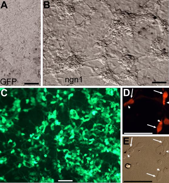
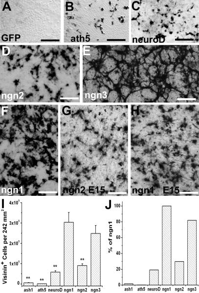
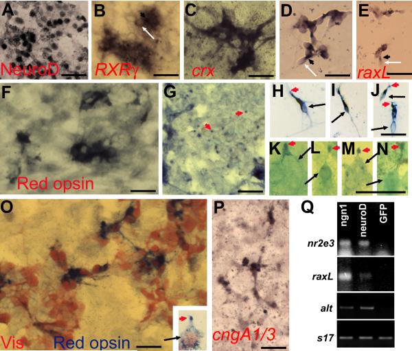
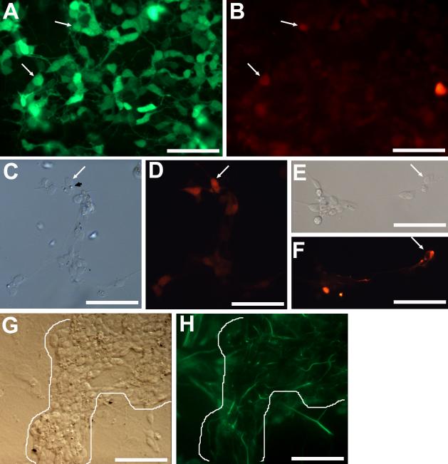
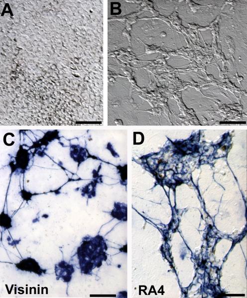
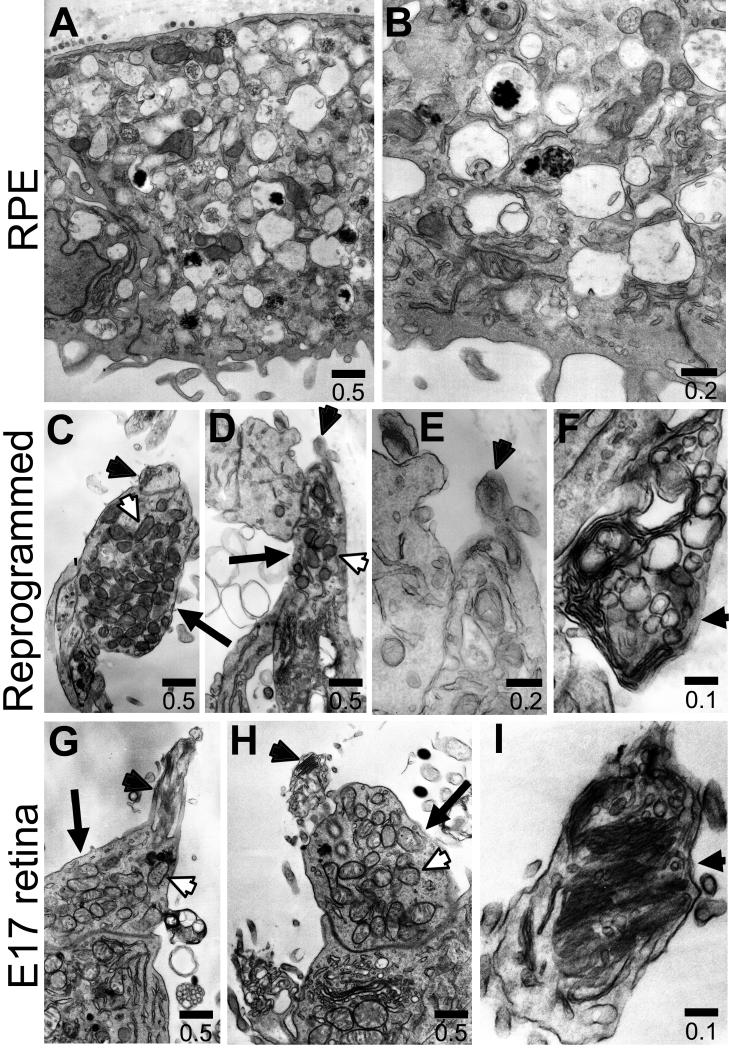
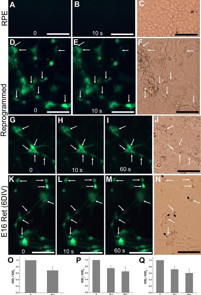
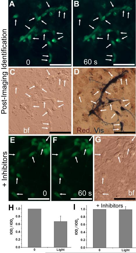
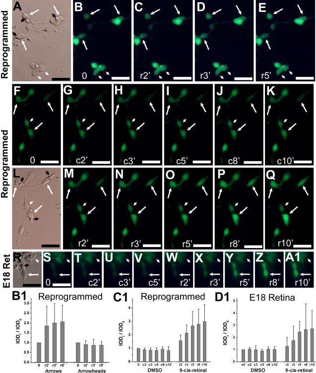
Photoreceptors are highly specialized sensory neurons in the retina, and their degeneration results in blindness. Replacement with developing photoreceptor cells promises to be an effective therapy, but it requires a supply of new photoreceptors, because the neural retina in human eyes lacks regeneration capability. We report efficient generation of differentiating, photoreceptor-like neurons from chick retinal pigment epithelial (RPE) cells propagated in culture through reprogramming with neurogenin1 (ngn1). In reprogrammed culture, a large number of the cells (85.0% +/- 5.9%) began to differentiate toward photoreceptors. Reprogrammed cells expressed transcription factors that set in motion photoreceptor differentiation, including Crx, Nr2E3, NeuroD, and RXRgamma, and phototransduction pathway components, including transducin, cGMP-gated channel, and red opsin of cone photoreceptors (equivalent to rhodopsin of rod photoreceptors). They developed inner segments rich in mitochondria. Furthermore, they responded to light by decreasing their cellular free calcium (Ca(2+)) levels and responded to 9-cis-retinal by increasing their Ca(2+) levels after photobleaching, hallmarks of photoreceptor physiology. The high efficiency and the advanced photoreceptor differentiation indicate ngn1 as a gene of choice to reprogram RPE progeny cells to differentiate into photoreceptor neurons in future cell replacement studies.


Figures

Fig. 1
Fig. 1
RPE cell cultures reprogrammed by ngn1. A,B: Bright-field views of a control culture infected with retrovirus RCAS-GFP (A) and a reprogrammed culture infected with RCAS-ngn1 (B). Red asterisks (*) in B mark cells clusters, which are absent in the control. C: Epi-fluorescence of visinin immunostaining of a ngn1-reprogrammed culture. D,E: Morphology of visinin+ cells viewed with bright-field (E) and epi-fluorescence (D). Arrows point to the cell body, and arrowheads point to a structural feature reminiscent of the lipid-droplet typically present in chick photoreceptors. Scale bars: 50 μm. A magenta-green copy is available as supplementary data.
Fig. 2
Fig. 2
Prevalence of visinin+ cells in RPE cell cultures subjected to reprogramming by various factors/genes. A-F: Representative images of immunodetection of visinin in E6 RPE cell cultures infected with retrovirus RCAS expressing GFP (A; a negative control), ath5 (B), neuroD (C), ngn2 (D), ngn3 (E), and ngn1 (F). G,H: Immunostaining for visinin in an E15 RPE cell culture infected with RCAS-ngn2 (G) or RCAS-ngn1 (H). I: Calculated numbers of visinin+ cells per 242 mm2 area in E6 RPE cell cultures reprogrammed with the different genes as shown. J: Calculated percentage of visinin+ cells in E6 RPE cell cultures reprogrammed with the different genes as shown against ngn1-reprogrmaed culture. Scale bars: 100 μm.
Fig. 3
Fig. 3
Expression of photoreceptor-specific genes in RPE cell cultures infected with RCAS-ngn1. A-E: Induction of transcription factors important for photoreceptor differentiation in ngn1-reprogrammed cultures. A: Immunostaining for NeuroD. B: In situ hybridization for RXRγ mRNA. C,D: In situ hybridization for crx mRNA in primary culture (C) and in re-seeded culture (D). E: In situ hybridization for raxL mRNA. F-K: Expression of phototransduction components in ngn1-reprogrammed cultures. F,G: Anti-red opsin immunostaining of the cell body (F) and dot-like structures at places (of the culture) where more pigmented RPE cells were present (G). H-N: Morphologies of red opsin+ cells in a re-seeded culture under a 40x objective (H-J) or in a primary culture under a 100x objective (K-N). Arrows point to cell bodies. Arrowheads point to cells’ apices decorated by anti-red opsin immunostaining. O: Double-staining with antibodies against visinin (in red) and red opsin (in blue). Inset: A higher magnification view of a double-labeled cell showing its cell body (arrow) and its apex decorated by anti-red opsin (arrowhead). P: In situ hybridization detection of the α-subunits of cone and rod CNG channels (cngA1/3). Q: RT-PCR detection of the expression of two transcription factors (nr2e3 and raxL), one phototransduction component (α-transducin, alt), and a control gene (s17) in cultures expressing RCAS-ngn1, RCAS-neuroD, or RCAS-GFP. Scale bars: 25 μm. A magenta-green copy is available as supplementary data.
Fig. 4
Fig. 4
Detection of markers of various retinal cell types in ngn1-reprogrammed cultures. A,B: Double-labeling for visinin (A) and calretinin (B) in a primary, reprogrammed culture. Arrows point to double-labeled cells. C,D: Morphologies of calretinin+ cells in a reseeded culture, viewed with bright field (C) or epi-fluorescence (D). The arrow points to a calretinin+ cell displaying a lipid-droplet-like structure. E,F: An RA4+ cell (arrow) with a long process in a reseeded culture viewed with bright field (E) or epi-fluorescence (F). G,H: Vimentin+ cells in a primary, reprogrammed culture viewed with bright field (G) or epi-fluorescence (H). Note: Long processes that were immuno-positive were present only at places occupied by cell clusters (outlined). Scale bars: 50 μm. A magenta-green copy is available as supplementary data.
Fig. 5
Fig. 5
Ngn3-reprogrammed E6 RPE cell cultures. A,B: Bright-field views of a control culture infected with RCAS-GFP (A) and a culture infected by RCAS-ngn3 (B). C,D: Anti-visinin (C) and RA4 (D) immunostaining of cultures infected with RCAS-ngn3. Scale bars: 50 μm.
Fig. 6
Fig. 6
Ultrastructures examined with electron micrography. A-B: Cells in a control RPE cell culture. C-F: ngn1-reprogrammed cells. D-I: Developing photoreceptors in E17 chick retina. Arrows: Inner segment; arrowheads: membranous expansion atop inner segments; white arrowheads: mitochondria. The scale bars are in μm.
Fig. 7
Fig. 7
Light responses examined with Ca2+ imaging. A-C: Images before (A) and after 10 seconds (10 s, B) of light exposure of a control RPE cell culture infected with RCAS. D-J: Images of RPE cell cultures infected with RCAS-ngn1 before (D,G) and after 10 seconds (E,H) and 60 seconds (I) of light exposure. K-N: Images of E16 chick retinal cells after 6 days in culture [E16 Ret (6DIV)]. C,F,J,N: Bright field images. Arrows: cells with noticeable reductions in fluorescence intensities. Arrowheads: lipid-droplet-like structures. O-Q: Calculated IOD ratios (IODt / IOD0) shown as means and SDs of 13-15 cells including those within the image of D,E, G-I, K-M, respectively. Scale bars: 50 μm.
Fig. 8
Fig. 8
Post-Ca2+ imaging immunostaining and effect of CNG channel blockers on light response. A-D: Double immunocytochemistry with antibodies against red opsin and visinin post Ca2+ imaging. Shown are images before (A) and after 60 seconds (B) of light exposure, a bright field view (C), and after double-immunostaining (D). Arrows point to cells with noticeable decreases in fluorescence intensities and positive for visinin (Vis, in blue) and/or red opsin (Red, in red). E-G: Images before (E) and after 60 seconds (F) of light exposure in the presence of CNG channel blockers dichlorobenzamil and l-cis-diltiazem (3 μM each). Arrows point to cells with photoreceptor-like morphologies and without significant reduction in their fluorescence intensities. H,I: Calculated IOD ratios (IODt / IOD0) shown as means and SDs of 14 and 10 cells including those in images A,B and E,F, respectively. Scale bars: 50 μm.
Fig. 9
Fig. 9
Response to 9-cis-retinal examined with Ca2+ imaging. A-E: Images of reprogrammed cells (in a reseeded culture) after light bleaching (B) and at 2 (C), 3 (D), and 5 (E) minutes after administration of 9-cis-retinal (r2′, r3′, r5′). F-Q: Images of reprogrammed cells after light bleaching (F) and at the indicated number of minutes (2′-10′) after sequential administration of vehicle control (c, G-K), replacement of medium (L), and then 9-cis-retinal (r, M-Q). R-A1: images of photoreceptor cells in an E18 retinal cell culture after 6 DIV. Arrows: cells showing increases in fluorescence intensity. White arrowheads: cells lacking such an increase. Black arrowheads: lipid droplet-like structure. B1-D1: The calculated IOD ratios (IODt / IOD0) were shown as means and SDs of 4-8 cells from experiments B-E, F-Q, and S-A1, respectively. A,L,R: Bright field images. Scale bars: 20 μm.

Similar articles

Cited by

References

    1. Adler R, Curcio C, Hicks D, Price D, Wong F. Cell death in age-related macular degeneration. Mol Vis. 1999;5:31. - PubMed
    1. Adler R, Lindsey JD, Elsner CL. Expression of cone-like properties by chick embryo neural retina cells in glial-free monolayer cultures. J Cell Biol. 1984;99:1173–1178. - PMC - PubMed
    1. Ahmad I, Tang L, Pham H. Identification of neural progenitors in the adult mammalian eye. Biochem Biophys Res Commun. 2000;270:517–521. - PubMed
    1. Aramant RB, Seiler MJ. Retinal transplantation--advantages of intact fetal sheets. Prog Retin Eye Res. 2002;21:57–73. - PubMed
    1. Austin CP, Feldman DE, Ida JA, Cepko CL. Vertebrate retinal ganglion cells are selected from competent progenitors by the action of Notch. Development. 1995;121:3637–3650. - PubMed

Publication types

MeSH terms