Case Reports
. 2009 Dec 23:10:155.
doi: 10.1186/1471-2202-10-155.

A case of polymicrogyria in macaque monkey: impact on anatomy and function of the motor system

Affiliations
Case Reports

A case of polymicrogyria in macaque monkey: impact on anatomy and function of the motor system

Eric Schmidlin et al. BMC Neurosci. .

Abstract

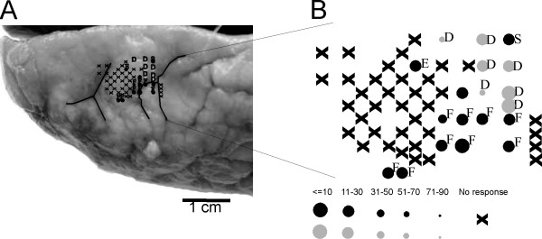
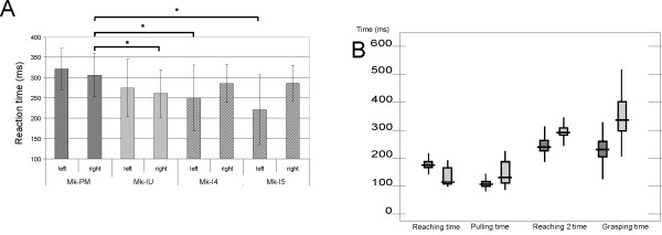
Background: Polymicrogyria is a malformation of the cerebral cortex often resulting in epilepsy or mental retardation. It remains unclear whether this pathology affects the structure and function of the corticospinal (CS) system. The anatomy and histology of the brain of one macaque monkey exhibiting a spontaneous polymicrogyria (PMG monkey) were examined and compared to the brain of normal monkeys. The CS tract was labelled by injecting a neuronal tracer (BDA) unilaterally in a region where low intensity electrical microstimulation elicited contralateral hand movements (presumably the primary motor cortex in the PMG monkey).

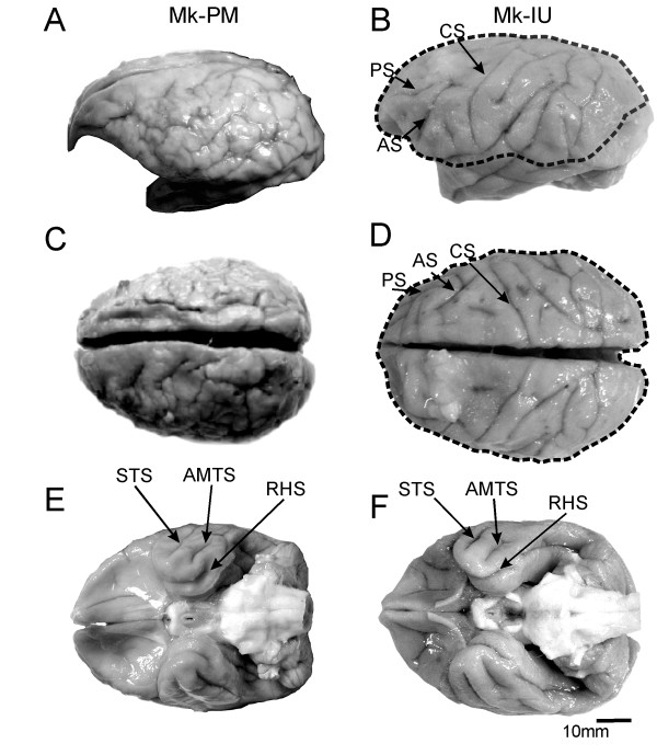
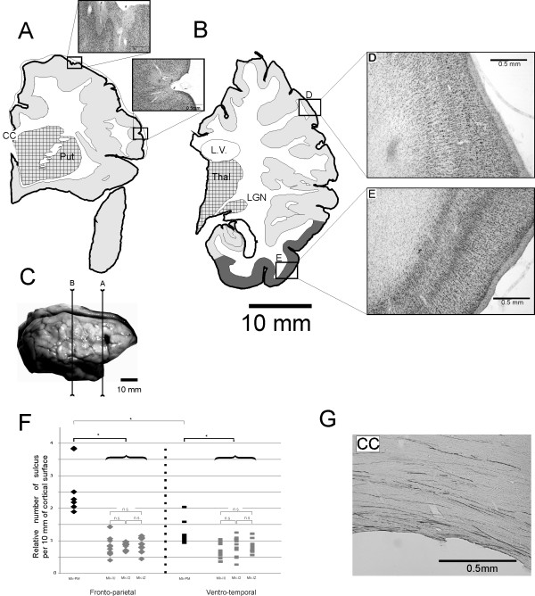
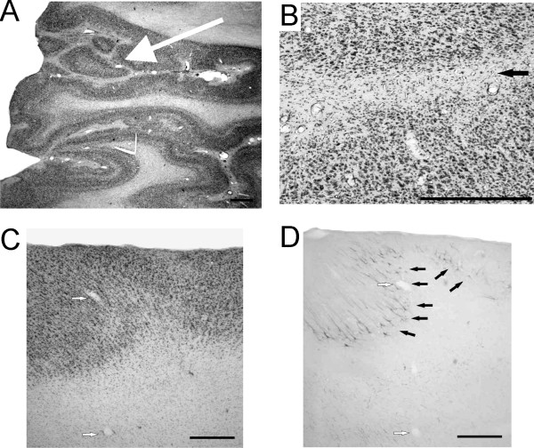
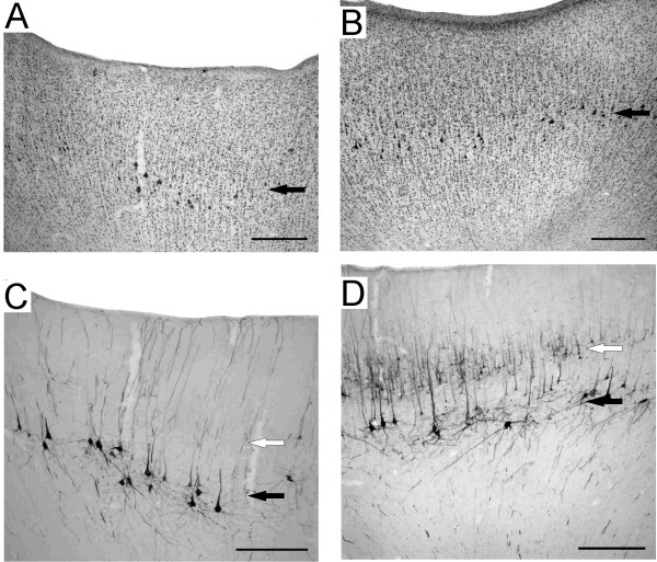
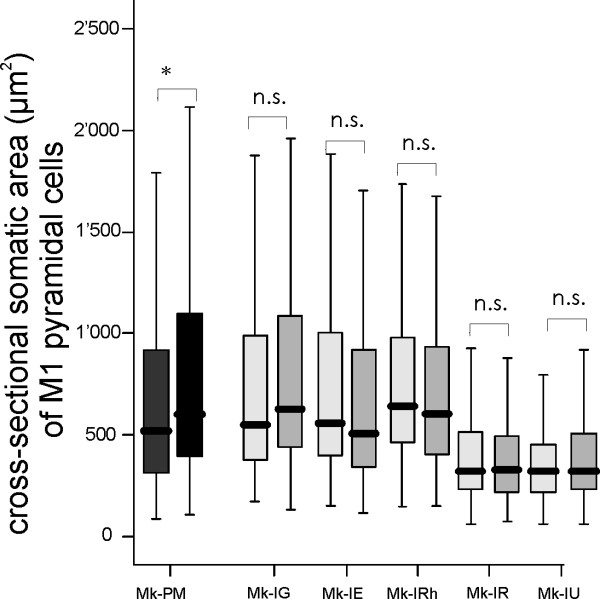
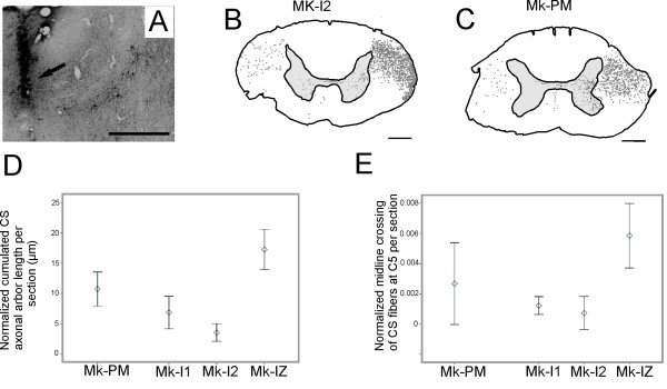
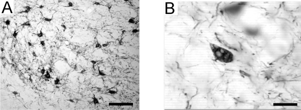
Results: The examination of the brain showed a large number of microgyri at macro- and microscopic levels, covering mainly the frontoparietal regions. The layered cortical organization was locally disrupted and the number of SMI-32 stained pyramidal neurons in the cortical layer III of the presumed motor cortex was reduced. We compared the distribution of labelled CS axons in the PMG monkey at spinal cervical level C5. The cumulated length of CS axon arbors in the spinal grey matter was not significantly different in the PMG monkey. In the red nucleus, numerous neurons presented large vesicles. We also assessed its motor performances by comparing its capacity to execute a complex reach and grasp behavioral task. The PMG monkey exhibited an increase of reaction time without any modification of other motor parameters, an observation in line with a normal CS tract organisation.

Conclusion: In spite of substantial cortical malformations in the frontal and parietal lobes, the PMG monkey exhibits surprisingly normal structure and function of the corticospinal system.


Figures

Figure 1
Figure 1
macroscopic views of the brain. Macroscopic appearance of the brain of the PMG monkey Mk-PM (A, C, E) and of a normal monkey Mk-IU (B, D, F) in lateral (A and B), dorsal (C and D) and ventral (E and F) views. Comparison of panels A and C with B and D shows a clear excessive number of small gyri in Mk-PM, and a loss of the normal topography of the brain, such as the disappearance of the normally well defined arcuate (AS), central (CS) or principal (PS) sulci. Nevertheless, the topographical organization of the ventral part of the PMG brain (E) is closer to that of the normal brain (F), as the superior temporal (STS), the anterior middle temporal (AMTS) or the rhinal sulci (RHS) are clearly identifiable. The extent of the cortical malformation in Mk-PM was projected over the healthy brain of Mk-IU (dashed line).
Figure 2
Figure 2
Reconstructions of frontal Nissl-stained sections in PMG monkey. Panels A and B: Location of cortical malformation on coronal sections in the right hemisphere at two rostrocaudal levels. Sub-cortical structures, such as the thalamus (Thal.), the lateral geniculate nucleus (LGN), the putamen (Put) or the corpus callosum (CC) are visible and they do not present conspicuous abnormalities, but the lateral ventricle (L.V.) is enlarged, including two enlarged photo micrograph of the cortex showing the microscopic cortical ectopic sulci (#), where the layer I is clearly distinguishable. Light grey: unlayered cortex. Dark grey: normally layered cortex: Squared: subcortical nuclei Panel C: Photograph of the cerebral cortex in Mk-PM with the position of the sections depicted in panels A and B. Panels D and E: The cortical organization in layers is lost in the parietal lobe (D) but normal in the inferior temporal lobe (E). Panel F: Diagram showing the distribution of the number of sulci per cortical length (number of sulcus per 10 mm) in the PMG monkey (black) and in the three control monkeys, Mk-IU, Mk-I2 and Mk-IZ (grey). Diamonds represent the results obtained in the frontoparietal region and bars in the ventro-temporal region. N.s is for non-statistically significant. Panel G: Photomicrograph of the Corpus Callosum (CC) in the right hemisphere of Mk-PM showing large quantity of BDA stained fibers. Scale bar: 500 microns.
Figure 3
Figure 3
Abnormalities in cortical organization. Panel A: Photomicrograph of a Nissl stained section demonstrating the disorganization of the microgyrial cortex. The layered organization is irregular and anomalous, with the formation of small islets (arrow). Panel B: Photomicrograph showing the lack of lumen and of pia between two facing portions of cortex in a sulcus, resulting in the fusion of both layers I in a unique strip of white matter (black arrow). Panel C: Photomicrograph of a Nissl stained section showing the absence of well defined laminar organization. Panel D: Photomicrograph of a SMI-32 stained section adjacent to the section presented in panel C. Note that the SMI-32 positive neurons occupy positions in the cortex which vary strongly with regard of its distance to the cortex surface (black arrows). White arrows in panels C and D indicate the same blood vessels. Scale bars: 500 microns.
Figure 4
Figure 4
Cytoarchitecture of the micro-excitable cortex. Photomicrographs of motor cortex of the right hemisphere in the PMG monkey (A: Nissl staining; C: SMI-32 staining) and in the normal monkey (B: Nissl staining; D: SMI-32 staining). Black arrows show the location of pyramidal cells in layer V, whereas the open arrows show the location of layer III. Note the absence of layer III SMI-32 stained neurons in Mk-PM. Scale bars: 500 microns.
Figure 5
Figure 5
Somatic cross-sectional areas of SMI-32 positive neurons in layer V in motor cortex. Box and whisker plots showing the distribution of somatic cross-sectional areas of SMI-32 positive neurons in layer V in motor cortex for the PMG monkey (dark grey) and four normal monkeys (light grey). In the box and whisker plots, the thick horizontal line in the box corresponds to the median value, whereas the top and bottom of the box are for the 75 and 25 percentile values respectively. The top and bottom extremities of the vertical lines on each side of the box are for the 90 and 10 percentile values, respectively. Mk-PM exhibited a significant inter-hemispheric difference of cross-sectional soma area (*p < 0.0001; Mann and Whitney test) whereas, in the normal monkeys, the difference was not statistically significant (n.s. p > 0.05; Mann and Whitney test). For each monkey, the left box and whisker plot corresponds to data from the left hemisphere.
Figure 6
Figure 6
Corticospinal projections in PMG monkey. Panel A: Site of BDA injection in M1 hand area of the left hemisphere (arrow), close to identified layer V pyramidal neurons. Panels B and C: Reconstructions of BDA stained corticospinal (CS) fibers in coronal sections of the cervical spinal cord at the C5 level, as a result of BDA injection in the right motor cortex of a normal monkey (B) and in the left motor cortex of the PMG monkey (C); scale bar 1 mm. For better visual comparison of both reconstructions, the reconstruction in panel C was drawn with the left spinal side on the right side of the drawing. Grey dots indicate the location and distribution of the CS axons in the white matter. In both monkeys, most fibers were found in the dorsolateral funiculus (DLF) contralateral to the injection site, the rest running along the dorsolateral and ventral funiculi ipsilateral to the injection site. In comparison to normal monkeys, slightly fewer CS fibers were found in the grey matter ipsilateral to the injection side in the PMG monkey. Panel D: Normalized cumulated axonal arbor length of corticospinal projections in the cervical grey matter in the PMG monkey (black) and in three normal monkeys (grey). Panel E: Number of midline crossing CS fibers at cervical level C5. The number of fibers crossing the midline was normalized by dividing it by the total number of labelled CS fibers present in the white matter (see methods for detail).
Figure 7
Figure 7
Abnormal vesicles in RNm neurons. SMI-32 staining of the red nucleus pars magnocellularis (RNm) in the PMG monkey. The general structure of the nucleus is comparable to that observed in normal monkeys (A), but abnormal accumulation of vesicles were observed in SMI-32 positive RNm neurons (B). Scale bars: (A) 200 μm; (B) 50 μm.
Figure 8
Figure 8
Functional organisation of M1 in the PMG monkey. Panel A: Left frontal lobe region of the brain of the PMG monkey with the map of the sites where intracortical microstimulation (ICMS) has been performed at a stereotaxic position corresponding to M1 in normal monkeys. Note the difficulty to identify the central sulcus and the precentral gyrus. Panel B: Map of the sites where ICMS has been performed, with indications on the body part of the observed movements and of the minimal current required to elicit the movement. D: digit, F: face, E: elbow and S: shoulder. X: No response at 80 microamps of stimulation intensity.
Figure 9
Figure 9
Behavior: reach and grasp drawer task. Panel A: Reaction times (RT) for each hands in the behavioral unimanual tasks for the 4 monkeys involved in the behavioral reach and grasp drawer task. In each case, the hand with which the PMG monkey (Mk-PM) exhibited the shortest average RTs (right) has been statistically tested against the fastest hand of the 3 normal monkeys (Mk-IU, Mk-I4, Mk-I5) using a Mann-Whitney test. Panel B: Movement times corresponding to the following movement components: First reaching time: from 'hand movement onset' to 'touch knob', Pulling time: from 'touch knob' to 'drawer fully open', Second reaching time: from 'grasping hand movement onset' to 'enter drawer's well', Grasping time: from 'hand penetrating in the well' to 'hand out of the well'.

Similar articles

Cited by

References

    1. Crome L. Microgyria. J Pathol Bacteriol. 1952;64:479–495. doi: 10.1002/path.1700640308. - DOI - PubMed
    1. Galaburda AM, Sherman GF, Rosen GD, Aboitiz F, Geschwind N. Developmental dyslexia: four consecutive patients with cortical anomalies. Ann Neurol. 1985;18:222–233. doi: 10.1002/ana.410180210. - DOI - PubMed
    1. Guerrini R, Dravet C, Raybaud C, Roger J, Bureau M, Battaglia A, Livet MO, Colicchio G, Robain O. Neurological findings and seizure outcome in children with bilateral opercular macrogyric-like changes detected by MRI. Dev Med Child Neurol. 1992;34:694–705. - PubMed
    1. Chang BS, Piao X, Giannini C, Cascino GD, Scheffer I, Woods CG, Topcu M, Tezcan K, Bodell A, Leventer RJ, Barkovich AJ, Grant PE, Walsh CA. Bilateral generalized polymicrogyria (BGP): a distinct syndrome of cortical malformation. Neurology. 2004;62:1722–1728. - PubMed
    1. Clark M, Pitt M, Neville BG. Lower motor neuron involvement in perisylvian polymicrogyria. Dev Med Child Neurol. 2006;48:842–846. doi: 10.1017/S0012162206001800. - DOI - PubMed

Publication types

MeSH terms