. 2009 Dec 24:9:271.
doi: 10.1186/1471-2180-9-271.

Downregulation of protein kinase C-alpha enhances intracellular survival of Mycobacteria: role of PknG

Affiliations

Downregulation of protein kinase C-alpha enhances intracellular survival of Mycobacteria: role of PknG

Shivendra K Chaurasiya et al. BMC Microbiol. .

Abstract

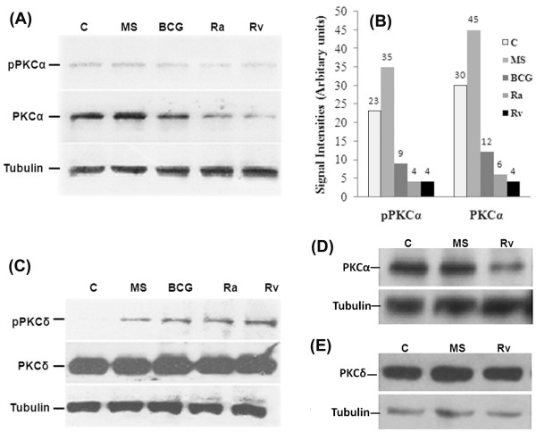
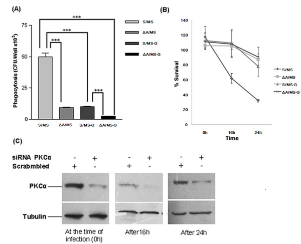
Background: Intracellular trafficking of mycobacteria is comprehensively dependent on the unusual regulation of host proteins. Recently, we have reported that infection of macrophages by Mycobacterium tuberculosis H37Rv (Rv) selectively downregulates the expression of PKCalpha while infection by Mycobacterium smegmatis (MS) does not.

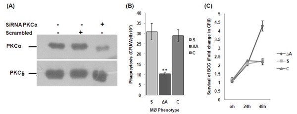
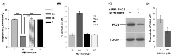
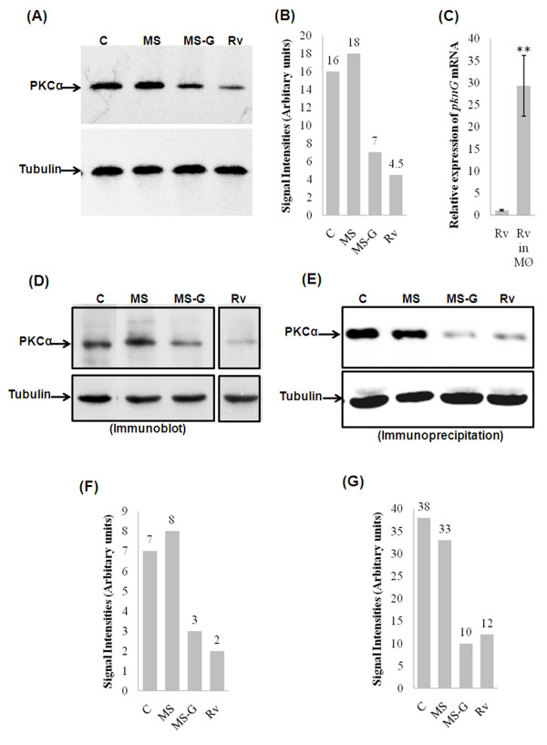
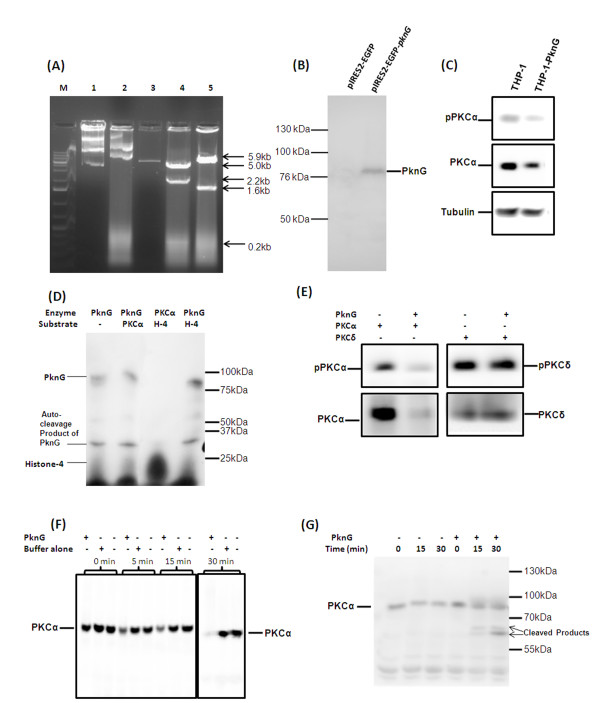
Results: Based on our earlier study, we have extrapolated for the first time that knockdown of PKCalpha, impairs phagocytosis of mycobacteria by macrophages while their intracellular survival is drastically increased. Mycobacterium bovis BCG (BCG) and Mycobacterium tuberculosis H37Ra (Ra) have also been shown to downregulate the expression of PKCalpha during the infection. Since PknG is uniquely expressed in BCG, Ra, Rv but not in MS and has been reported to promote intracellular survival of mycobacteria, led us to believe that PknG may be involved in such downregulation of PKCalpha. THP-1 cells infected with recombinant MS expressing PknG (MS-G), showed significant reduction in PKCalpha expression. In normal THP-1 cells survival of MS-G was enhanced as compared to MS, while their behavior in PKCalpha deficient cells could not be distinguished. The results strongly demonstrate that pathogenic mycobacteria recognize and then inhibit PKCalpha to circumvent phagocytosis and the hostile environment of macrophages. We emphasize that, this inhibition is controlled by PknG.

Conclusions: All together, our data reveal a mechanism that shows substantial interdependence of PKCalpha with PknG, in sustaining mycobacterial infection.


Figures

Figure 1
Figure 1
Downregulation of PKC-α expression by mycobacteria. THP-1 cells were incubated for 4 h in the presence of mycobacteria (MOI = 1:20) as indicated (C, uninfetced). The cells were lysed, and equal amounts of total cell lysates (20 μg) were resolved by SDS-PAGE and immunoblotted with an antibody against (A) PKC-α and phosphorylated form of PKC-α (Thr638), (B) Densitometric analysis of PKC-α and pPKC-α blots shown in fig. 1A, (C) PKC-δ and phospho-PKCδ (Thr505). The lower parts of the blots were probed with an anti-tubulin antibody, to assure equal protein loading (lower panel), (D) and (E) level of PKC-α and PKC-δ in mouse peritoneal macrophages. Each experiment was repeated at least 3 times.
Figure 2
Figure 2
Phagocytosis and survival of BCG in PKC-α deficient THP-1 cells. THP-1 cells were incubated in the presence of 30 nM PMA for 24 h. Then cells were transfected with 20 nM SiRNA and level of PKC-α were determined by immunoblotting. (A) 24 h after transfection, level of PKC-α and PKC-δ in cells transfected with SiRNA targeting PKC-α or scrambled SiRNA, (B) 24 h after transfection, (ΔA) cells transfected with SiRNA targeting PKC-α and (S) cells transfected with scrambled SiRNA and control cells (C) were infected with BCG (MOI = 1:10) for 2 h, washed and remaining extracellular bacilli were killed by amikacin treatment for 1 h and lysed in 0.05% SDS and plated. Colony forming units (cfu) were determined after 4 week of incubation. Tukey (T) test was performed for statistical analysis of data (C) Survival of BCG in THP-1 cells transfected with either SiRNA targeting PKC-α (ΔA) or scrambled SiRNA (S) after 24 and 48 h, since phagocytosis of BCG in control and PKC-α deficient cells was different, CFU at 0 h was considered 1 and survival of BCG is presented as fold increase in the number of cfu as compared to the initial phagocytosis. Data are means ± standard deviations from three independent experiments each performed in 4 replicates. (** = p < 0.005).
Figure 3
Figure 3
Phagocytosis and survival of MS in PKC-α deficient THP-1 cells. THP-1 cells were incubated in the presence of 30 nM PMA for 24 h. Then cells were transfected with 20 nM SiRNA and after 24 h level of PKC were determined by immunoblotting. (A) 24 h after transfection control cells (C) and (ΔA) cells transfected with SiRNA PKCα, (ΔD) cells transfected with SiRNA PKCδ, (S) cells transfected with scrambled SiRNA (PKC-α SiRNA which does not block PKCα), were infected with MS (MOI = 1:10) for 2 h, washed and remaining extracellular bacilli were killed by amikacin treatment for 1 h, again washed, lysed in 0.05% SDS and plated for cfu. 'T' test was performed for statistical analysis of data, (B) 24 h after infection % survival of MS in THP-1 cells transfected with either SiRNA targeting PKC-α (ΔA) or scrambled SiRNA (S), because phagocytosis of MS was different in control and PKC-α deficient cells, cfu at 0 h was considered 100% and survival of MS is presented as percentage of the initial cfu that survive in macrophages after 24 h. (C) 24 h after transfection, level of PKC-δ in cells transfected with SiRNA targeting PKC-δ or scrambled SiRNA, (D) Phagocytosis of MS by mouse macrophage cell line J774A.1 cells pretreated with an inhibitor of PKC-α (Go6976) for 30 minute before infection. Data are means ± standard deviations from three independent experiments each performed in 4 replicates. (*** = p < 0.0001, * = p < 0.05).
Figure 4
Figure 4
Downregulation of expression of macrophage PKC-α by recombinant mycobacteria expressing PknG. (A) The THP-1 cells infected with either wild type or recombinant mycobacteria were lysed, and equal amounts of total cell lysates (20 μg) were resolved by SDS-PAGE and immunoblotted with an antibody against PKCα. The lower parts of the blots were probed with an anti-tubulin antibody, to assure equal protein loading, (B) Densitometric analysis of blots shown in fig. 5A, (C) THP-1 cells infected with Rv were osmotically lysed and bacteria were recovered by centrifugation and total bacterial RNA was isolated. Total RNA was also isolated from bacterial suspension in RPMI-1640 medium which was used for infection of THP-1 cells. RNA samples were treated with DNAse I and cDNA were prepared using random hexamer primers and was used as template for Cyber Green real time PCR using pknG specific primers (values presented are normalized against 16S rRNA), Data are means ± standard deviations from five independent experiments each performed in 3 replicates. (** = p < 0.005). (D) experiment identical to 5A was performed with J774A.1 cells, (E) equal amounts of total cell lysates of J774A.1 cells infected with mycobacteria were immunoprecipitated with anti-PKC-α antibody and level of PKC-α was analyzed by immunoblotting. Same amounts of lysates were also immunoprecipitated with anti-tubulin antibody to serve as control, (F) Densitometric analysis of blots shown in fig. 5D, (G) Densitometric analysis of blots shown in fig.5E. The experiments were repeated at least 3 times.
Figure 5
Figure 5
Comparison of phagocytosis and intracellular survival of MS and MS-G in normal and in PKC-α deficient THP-1 cells. (A) THP-1 cells were incubated in the presence of 30 nM PMA for 24 h. Cells were then transfected either with SiRNA targeting PKC-α (ΔA) or scrambled SiRNA (S) and after 24 h were infected with MS or MS-G (MOI = 1:10) for 2 h, washed and remaining extracellular bacilli were killed by amikacin treatment for 1 h, again washed and internalized bacteria were released by lysis of macrophages with 0.05% SDS and plated then cfu were counted, (S/MS) phagocytosis of MS by normal THP-1 cells, (ΔA/MS) phagocytosis of MS by PKC-α deficient THP-1 cells, (S/MS-G) phagocytosis of MS-G by normal THP-1 cells, (ΔA/MS-G) phagocytosis of MS-G by PKC-α deficient THP-1 cells. 'T' test was performed for statistical analysis of data. (B) % survival of MS and MS-G in normal and PKC-α deficient THP-1 cells. Because, phagocytosis of MS and MS-G were different in control and in PKC-α deficient cells, cfu at 0 h was considered 100% and survival of MS is presented as percentage of the initial cfu. (C) At each time point of experiment, level of PKC-α in cells transfected either with SiRNA targeting PKC-α or scrambled SiRNA was also determined by immunoblotting, to confirm the levels of PKC-α throughout the experiment. Data are means ± standard deviations from three independent experiments each performed in 4 replicates. (*** = p < 0.0001).
Figure 6
Figure 6
Mechanism of downregulation of PKC-α by PknG. (A) Cloning of pknG in pIRES2-EGFP vector; M, DNA ladder; 1, pIRES2-EGFP-pknG undigested; 2, pIRES2-EGFP undigested; 3, pIRES2-EGFP digested with BamHI; 4, pIRES2-EGFP-pknG digested with HindIII; 5, pIRES2-EGFP-pknG digested with BamHI, right oriented recombinants will produce 1.6 kb fragment; (B) and (C) pIRES2-EGFP-pknG was transfected in THP-1 cells and after 48 h cells were lysed and immunoblotted with anti-serum against PknG and with PKC-α antibodies, lane 1 macrophages transfected with vector alone and lane 2 transfected with pIRES2-EGFP-pknG. (D) 5 μg PknG was incubated with immunoprecipitated PKC-α in kinase buffer for 30 min in presence of [γ32P]-ATP then resolved by 8% SDS-PAGE and exposed to X-Ray film., lane 1 PknG alone; lane 2 PKC-α and PknG, lane 3 PKC-α and Histone-4 and lane 4 PknG and Histone-4. (E) THP-1 cell lysate was immunoprecipitated with either antibodies against PKC-α or PKC-δ using protein G Sepharose. The immunoprecipitated proteins were incubated with 5 μg purified PknG for 1 h and immunoblotted with PKC-α and PKC-δ antibodies. (F) Macrophage cell lysate (50 μg) was incubated with 5 μg purified PknG or buffer alone for indicated times and immunoblotted with PKC-α antibodies. (G) THP-1 cell lysate was immunoprecipitated with antibodies against PKC-α and immunoprecipitates were treated with 5 μg of purified PknG for the time points as indicated and then immunoblotted with PKC-α antibody. The experiments were repeated at least 3 times.

Similar articles

Cited by

References

    1. Koul A, Herget T, Klebl B, Ullrich A. Interplay between mycobacteria and host signalling pathways. Nat Rev Microbiol. 2004;2:189–202. doi: 10.1038/nrmicro840. - DOI - PubMed
    1. Malik ZA, Denning GM, Kusner DJ. Inhibition of ca2+ signaling by Mycobacterium tuberculosis is associated with reduced phagosome-lysosome fusion an d increased survival within human macrophages. J Exp Med. 2000;191:287–302. doi: 10.1084/jem.191.2.287. - DOI - PMC - PubMed
    1. Malik ZA, Iyer SS, Kusner DJ. Mycobacterium tuberculosis phagosomes exhibit altered calmodulin-dependent signal transduction: contribution to inhibition of phagosome-lysosome fusion and intracellular survival in human macrophages. J Immunol. 2001;166:3392–3401. - PubMed
    1. Rao KMK. MAP kinase activation in macrophages. J Leukoc Biol. 2001;69:3–10. - PubMed
    1. Cole ST, Brosch R, Parkhill J, Garnier T, Churcher C, Harris D, Gordon SV, Eiglmeier K, Gas S, Barry CE, Tekaia F, Badcock K, Basham D, Brown D, Chillingworth T, Connor T, Davies R, Devlin K, Feltwell T, Gentles S, Hamlin N, Holroyd S, Hornsby T, Jagels K, Krogh A, McLean J, Moule S, Murphy L, Oliver K, Osborne J, Quail MA, Rajandream MA, Rogers J, Rutter S, Seeger K, Skelton J, Squares R, Squares S, Sulston JE, Taylor K, Whitehead, Barrell BG. Deciphering the biology of Mycobacterium tuberculosis from the complete genome sequence. Nature. 1998;393:537–544. doi: 10.1038/31159. - DOI - PubMed

Publication types

MeSH terms

Substances

LinkOut - more resources