

Channel catfish soluble FcmuR binds conserved linear epitopes present on Cmu3 and Cmu4
- PMID: 20031218
- PMCID: PMC2830281
- DOI: 10.1016/j.molimm.2009.11.026
Channel catfish soluble FcmuR binds conserved linear epitopes present on Cmu3 and Cmu4
Abstract
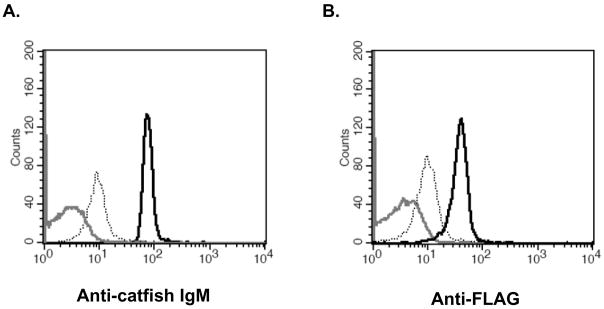
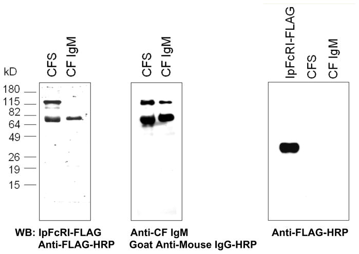
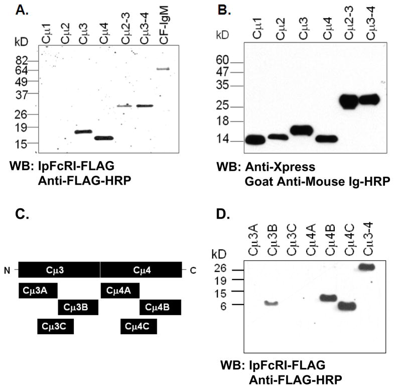
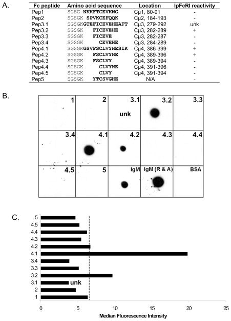
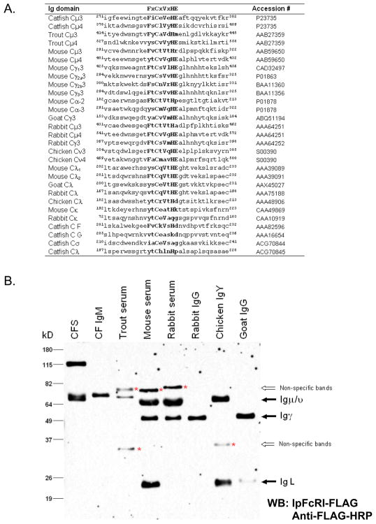
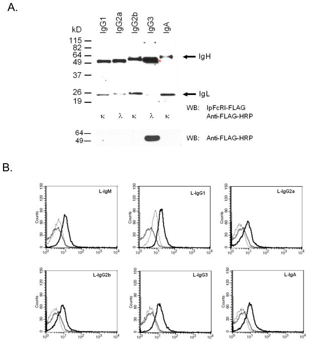
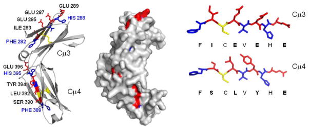
A linear epitope on catfish IgM has been identified as the docking site for the catfish soluble FcmuR (IpFcRI). Western blot analyses and latex bead binding assays identified the consensus octapeptide motif FxCxVxHE located at the second cysteine that forms the intrachain disulfide bond of the catfish Cmu3 and Cmu4 immunolglobulin (Ig) domains as the IpFcRI binding sites. Furthermore, molecular modeling of catfish Cmu3 and Cmu4 confirmed that the octapeptide in both of these domains is accessible for IpFcRI interactions. In addition, since this octapeptide motif is also found in other vertebrate Ig domains, IpFcRI binding to Ig heavy (H) and light (L) chains from rainbow trout, chicken, mouse, rabbit, and goat were examined by Western blot analyses and latex bead binding assays. IpFcRI readily bound reduced rainbow trout (Igmu), chicken (Ignu), mouse (Igmu, Iggamma1, Iggamma2a, Iggamma2b, and Igalpha), rabbit (Igmu and Iggamma) and goat (Iggamma) IgH chains, and mouse Igkappa and Iglambda, and chicken Iglambda IgL chains. IpFcRI also bound mouse IgM, IgA and IgG subclasses when examined under native conditions.
Copyright 2009 Elsevier Ltd. All rights reserved.
Figures
Similar articles
-
Identification and characterization of a FcR homolog in an ectothermic vertebrate, the channel catfish (Ictalurus punctatus).J Immunol. 2006 Aug 15;177(4):2505-17. doi: 10.4049/jimmunol.177.4.2505. J Immunol. 2006. PMID: 16888012
-
Mapping of the binding site for FcμR in human IgM-Fc.Biochim Biophys Acta Proteins Proteom. 2020 Jan;1868(1):140266. doi: 10.1016/j.bbapap.2019.140266. Epub 2019 Aug 23. Biochim Biophys Acta Proteins Proteom. 2020. PMID: 31449905 Free PMC article.
-
Characterization of anti-channel catfish IgL sigma monoclonal antibodies.Vet Immunol Immunopathol. 2010 Jun 15;135(3-4):325-8. doi: 10.1016/j.vetimm.2010.01.004. Epub 2010 Jan 25. Vet Immunol Immunopathol. 2010. PMID: 20149930
-
Channel catfish immunoglobulins: repertoire and expression.Dev Comp Immunol. 2006;30(1-2):77-92. doi: 10.1016/j.dci.2005.06.016. Dev Comp Immunol. 2006. PMID: 16153707 Review.
-
Role of the IgM Fc Receptor in Immunity and Tolerance.Front Immunol. 2019 Mar 22;10:529. doi: 10.3389/fimmu.2019.00529. eCollection 2019. Front Immunol. 2019. PMID: 30967868 Free PMC article. Review.
Cited by
-
Functional Characterization of Largemouth Bass (Micropterus salmoides) Soluble FcγR Homolog in Response to Bacterial Infection.Int J Mol Sci. 2022 Nov 9;23(22):13788. doi: 10.3390/ijms232213788. Int J Mol Sci. 2022. PMID: 36430268 Free PMC article.
-
A soluble FcγR homolog inhibits IgM antibody production in ayu spleen cells.Zool Res. 2019 Sep 18;40(5):404-415. doi: 10.24272/j.issn.2095-8137.2019.056. Zool Res. 2019. PMID: 31343855 Free PMC article.
-
Site-Specific N-Glycan Characterization of Grass Carp Serum IgM.Front Immunol. 2018 Nov 14;9:2645. doi: 10.3389/fimmu.2018.02645. eCollection 2018. Front Immunol. 2018. PMID: 30487799 Free PMC article.
-
The structure of the teleost Immunoglobulin M core provides insights on polymeric antibody evolution, assembly, and function.Nat Commun. 2023 Nov 21;14(1):7583. doi: 10.1038/s41467-023-43240-z. Nat Commun. 2023. PMID: 37989996 Free PMC article.
References
-
- Armant M, Rubio M, Delespesse G, Sarfati M. Soluble CD23 directly activates monocytes to contribute to the antigen-independent stimulation of resting T cells. J Immunol. 1995;155:4868–75. - PubMed
-
- Astier A, de la Salle H, de la Salle C, Bieber T, Esposito-Farese ME, Freund M, Cazenave JP, Fridman WH, Teillaud JL, Hanau D. Human epidermal Langerhans cells secrete a soluble receptor for IgG (Fc gamma RII/CD32) that inhibits the binding of immune complexes to Fc gamma R+ cells. J Immunol. 1994;152:201–12. - PubMed
-
- Bengten E, Clem LW, Miller NW, Warr GW, Wilson M. Channel catfish immunoglobulins: repertoire and expression. Dev Comp Immunol. 2006a;30:77–92. - PubMed
-
- Bengten E, Quiniou S, Hikima J, Waldbieser G, Warr GW, Miller NW, Wilson M. Structure of the catfish IGH locus: analysis of the region including the single functional IGHM gene. Immunogenetics. 2006b;58:831–44. - PubMed
-
- Bengten E, Quiniou SM, Stuge TB, Katagiri T, Miller NW, Clem LW, Warr GW, Wilson M. The IgH locus of the channel catfish, Ictalurus punctatus, contains multiple constant region gene sequences: different genes encode heavy chains of membrane and secreted IgD. J Immunol. 2002;169:2488–97. - PubMed
Publication types
MeSH terms
Substances
Grants and funding
LinkOut - more resources
Full Text Sources
Molecular Biology Databases
Miscellaneous
