. 2009 Aug;2(4):371-8.
doi: 10.1161/CIRCGENETICS.108.832345. Epub 2009 May 15.

Cardiac resynchronization therapy corrects dyssynchrony-induced regional gene expression changes on a genomic level

Affiliations

Cardiac resynchronization therapy corrects dyssynchrony-induced regional gene expression changes on a genomic level

Andreas S Barth et al. Circ Cardiovasc Genet. 2009 Aug.

Abstract

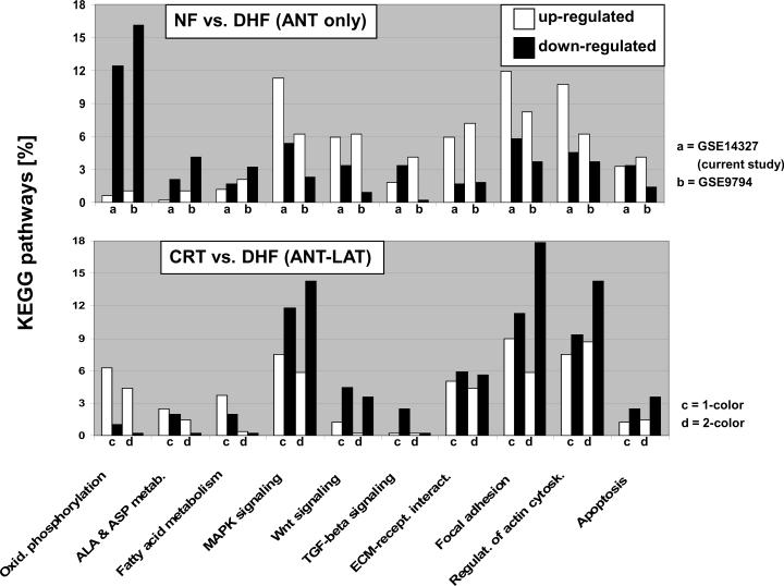
Background: Cardiac electromechanical dyssynchrony causes regional disparities in workload, oxygen consumption, and myocardial perfusion within the left ventricle. We hypothesized that such dyssynchrony also induces region-specific alterations in the myocardial transcriptome that are corrected by cardiac resynchronization therapy (CRT).

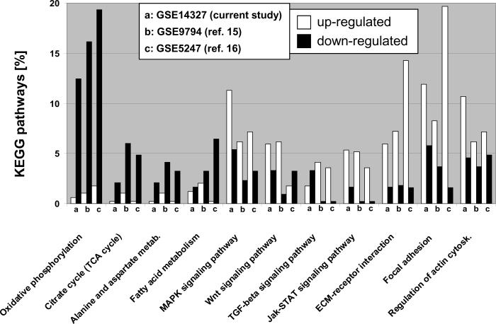
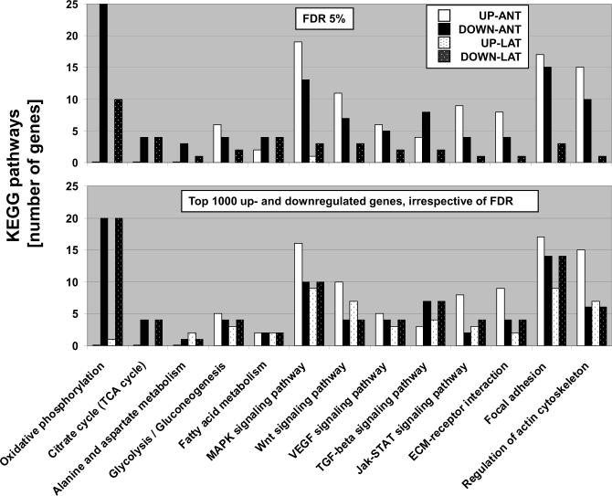
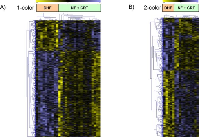
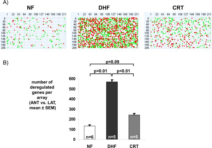
Methods and results: Adult dogs underwent left bundle branch ablation and right atrial pacing at 200 bpm for either 6 weeks (dyssynchronous heart failure, n=12) or 3 weeks, followed by 3 weeks of resynchronization by biventricular pacing at the same pacing rate (CRT, n=10). Control animals without left bundle branch block were not paced (n=13). At 6 weeks, RNA was isolated from the anterior and lateral left ventricular (LV) walls and hybridized onto canine-specific 44K microarrays. Echocardiographically, CRT led to a significant decrease in the dyssynchrony index, while dyssynchronous heart failure and CRT animals had a comparable degree of LV dysfunction. In dyssynchronous heart failure, changes in gene expression were primarily observed in the anterior LV, resulting in increased regional heterogeneity of gene expression within the LV. Dyssynchrony-induced expression changes in 1050 transcripts were reversed by CRT to levels of nonpaced hearts (false discovery rate <5%). CRT remodeled transcripts with metabolic and cell signaling function and greatly reduced regional heterogeneity of gene expression as compared with dyssynchronous heart failure.

Conclusions: Our results demonstrate a profound effect of electromechanical dyssynchrony on the regional cardiac transcriptome, causing gene expression changes primarily in the anterior LV wall. CRT corrected the alterations in gene expression in the anterior wall, supporting a global effect of biventricular pacing on the ventricular transcriptome that extends beyond the pacing site in the lateral wall.


Figures

Figure 1
Figure 1. KEGG pathway analysis in tachycardia-pacing induced heart failure
Transcripts up- and down-regulated by ventricular tachypacing in canine ventricular myocardium are represented by white and black columns, respectively and shown as percentage of eleven major KEGG pathways (studies a-c). Study (a) shows the comparison of anterior samples between NF and DHF hearts in the current Agilent-based microarray study. Studies (b) and (c) show the results of two publicly available Affymetrix microarray datasets (Gene Expression Omnibus accession numbers 9794 and 5247, respectively) comparing myocardial tissue derived from the anterior LV wall from non-paced animals to dogs that were tachypaced for at least 3 weeks. Normalized data was downloaded from Gene Expression Omnibus and analyzed using the same methods employed in this study (a). For all KEGG pathways shown, a p-value of <0.05 (Fisher's exact test implemented in the “FatiGO+” tool) was achieved in at least two of the three different canine tachypacing studies presented. It is evident that energy-deriving processes including oxidative phosphorylation and TCA cycle are greatly down-regulated in pacing-induced HF, while various cell signaling pathways and extracellular matrix components are up-regulated. The high concordance of disease-specific gene expression patterns across independent studies and different microarray platforms serves as an independent validation of our results and suggests a common disease-specific genomic fingerprint.
Figure 2
Figure 2. Regional KEGG pathway analysis in tachycardia-pacing induced heart failure
KEGG pathways are plotted as numbers of up- and down-regulated transcripts that were differentially expressed between non-failing and failing myocardium. Transcripts up- and down-regulated by ventricular tachypacing are represented by white and black columns (anterior wall), or textured white and black bars (lateral wall), respectively. The upper panel compares 11 KEGG pathways identified by SAM analysis in anterior and lateral myocardium, respectively. The small number of regulated genes found in lateral myocardium of DHF hearts limited the statistical power of the KEGG pathway analysis, thus an additional analysis was performed where KEGG pathways of the first 1000 up- and down-regulated transcripts were compared in both regions, irrespective of the significance level (lower panel). This approach was employed to determine if the changes in gene expression in the lateral wall differ only quantitatively from those in the anterior LV wall. The metabolic gene classes were still down-regulated; however, the pattern of gene expression for cell signaling pathways and extracellular matrix components differed considerably from the robust genomic fingerprint observed in anterior myocardium in DHF, suggesting also qualitatively different transcriptomic responses to electromechanical dyssynchrony in the anterior and lateral LV.
Figure 3
Figure 3. Clustering of regional differences between anterior and lateral myocardium
Unsupervised clustering of differentially expressed transcripts identified by SAM (multiclass, false discovery rate <5%) using Euclidean distance for one and two color microarray data (panel A and B, respectively) shows that transcript expression from CRT hearts cluster with NF hearts rather than with DHF samples. Each row represents data for one gene. The gene expression level is color-coded with yellow and blue representing low and high expression, respectively. For one-color data, the difference in gene expression between the anterior and lateral wall from the same heart was compared for NF, DHF and CRT animals.
Figure 4
Figure 4. Dyssynchrony leads to increased regional heterogeneity in gene expression that is partially reduced with CRT
(A) Pseudoimages of representative microarrays from NF, DHF and CRT hearts with 211 columns and 206 rows (44K array). RNA from the anterior and lateral regions was labeled with Cy3 and Cy5 and hybridized in a two-color design onto one array. Red and green dots represent statistically significant transcripts between anterior and lateral wall, respectively. (B) A bar plot of the number of deregulated genes comparing the anterior and lateral regions in NF, DHF and CRT hearts. In DHF, the number of differentially expressed transcripts between anterior and lateral wall increases 4-fold, while it is greatly reduced by CRT.
Figure 5
Figure 5. Partial correction of pacing-induced gene expression changes by CRT
A comparison of up- and down-regulated transcripts in KEGG pathways that were differentially expressed between non-failing and failing myocardium (upper panel) and between anterior and lateral LV myocardium (lower panel). Up- and down-regulated transcripts are represented by white and black columns, respectively. CRT partially restores dyssynchrony-induced gene expression changes in failing ventricular myocardium (up regulation of transcripts in oxidative phosphorylation pathways and down regulation of cell signaling pathways in the anterior wall).

Comment in

  • Pathways of the heart.
    Deo RC, Roth FP. Deo RC, et al. Circ Cardiovasc Genet. 2009 Aug;2(4):303-5. doi: 10.1161/CIRCGENETICS.109.892588. Circ Cardiovasc Genet. 2009. PMID: 20031600 Free PMC article. No abstract available.

References

    1. Rosamond W, Flegal K, Furie K, Go A, Greenlund K, Haase N, Hailpern SM, Ho M, Howard V, Kissela B, Kittner S, Lloyd-Jones D, McDermott M, Meigs J, Moy C, Nichol G, O'Donnell C, Roger V, Sorlie P, Steinberger J, Thom T, Wilson M, Hong Y. Heart disease and stroke statistics--2008 update: a report from the American Heart Association Statistics Committee and Stroke Statistics Subcommittee. Circulation. 2008;117:e25–146. - PubMed
    1. Baldasseroni S, Opasich C, Gorini M, Lucci D, Marchionni N, Marini M, Campana C, Perini G, Deorsola A, Masotti G, Tavazzi L, Maggioni AP. Left bundle-branch block is associated with increased 1-year sudden and total mortality rate in 5517 outpatients with congestive heart failure: a report from the Italian network on congestive heart failure. Am Heart J. 2002;143:398–405. - PubMed
    1. Bader H, Garrigue S, Lafitte S, Reuter S, Jais P, Haissaguerre M, Bonnet J, Clementy J, Roudaut R. Intra-left ventricular electromechanical asynchrony. A new independent predictor of severe cardiac events in heart failure patients. J Am Coll Cardiol. 2004;43:248–256. - PubMed
    1. Kass DA, Chen CH, Curry C, Talbot M, Berger R, Fetics B, Nevo E. Improved left ventricular mechanics from acute VDD pacing in patients with dilated cardiomyopathy and ventricular conduction delay. Circulation. 1999;99:1567–1573. - PubMed
    1. Spragg DD, Kass DA. Pathobiology of left ventricular dyssynchrony and resynchronization. Prog Cardiovasc Dis. 2006;49:26–41. - PubMed

Publication types

MeSH terms