. 2009 Dec;2(6):583-90.
doi: 10.1161/CIRCGENETICS.108.847814. Epub 2009 Sep 21.

In vivo genetic evidence for suppressing vascular and soft-tissue calcification through the reduction of serum phosphate levels, even in the presence of high serum calcium and 1,25-dihydroxyvitamin d levels

Affiliations

In vivo genetic evidence for suppressing vascular and soft-tissue calcification through the reduction of serum phosphate levels, even in the presence of high serum calcium and 1,25-dihydroxyvitamin d levels

Mutsuko Ohnishi et al. Circ Cardiovasc Genet. 2009 Dec.

Abstract

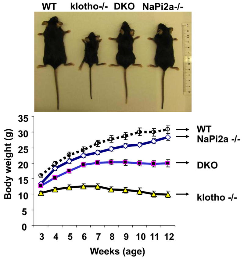
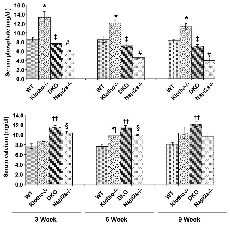
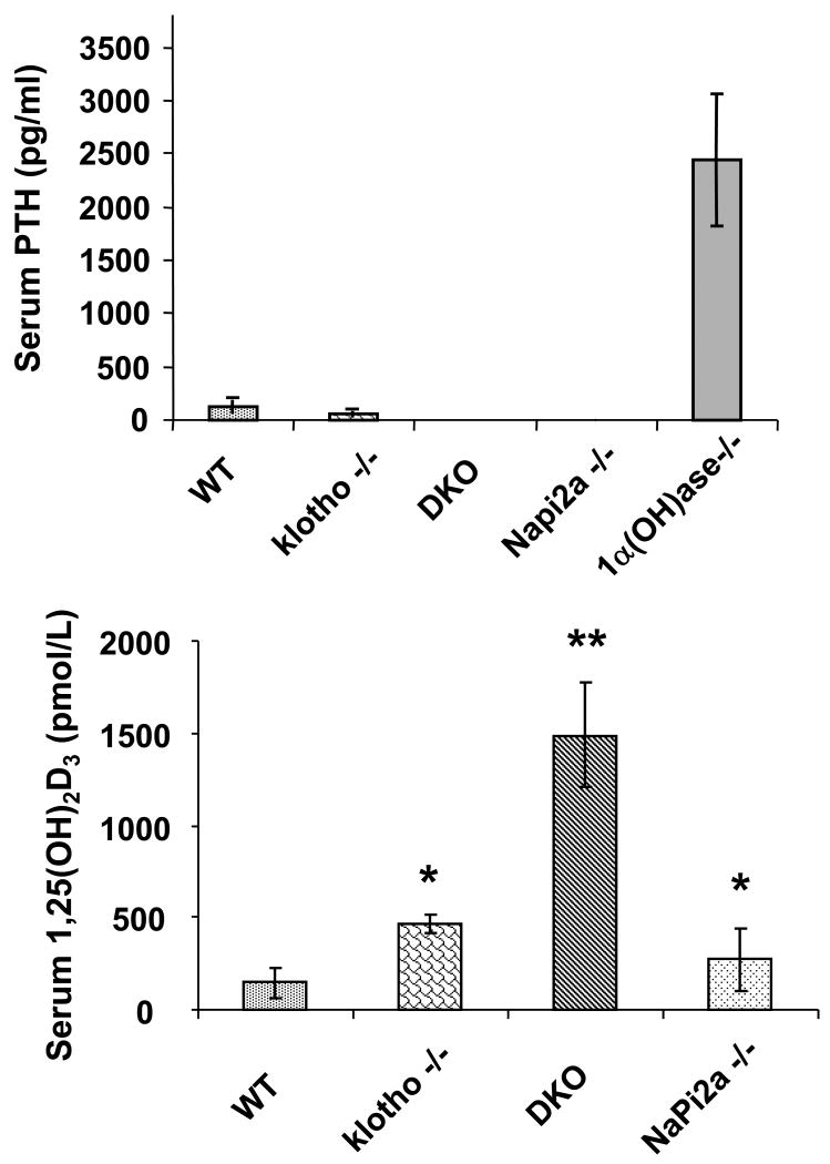
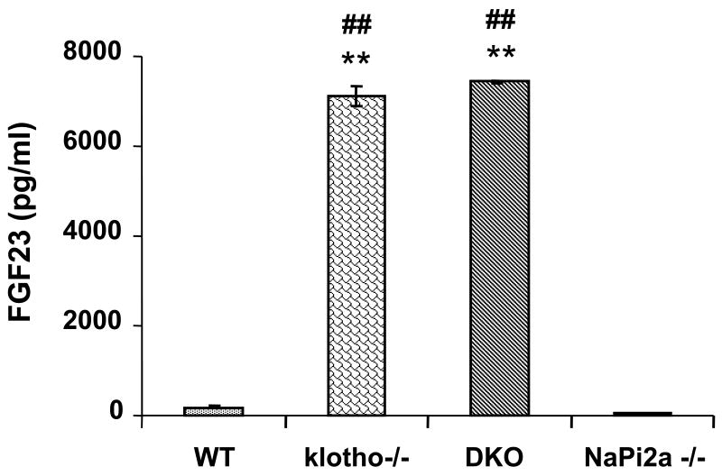
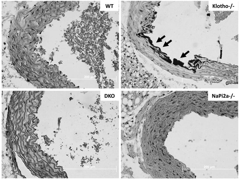
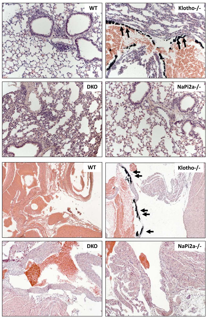
Background: Klotho-knockout mice (klotho(-/-)) have increased renal expression of sodium/phosphate cotransporters (NaPi2a), associated with severe hyperphosphatemia. Such serum biochemical changes in klotho(-/-) mice lead to extensive soft-tissue anomalies and vascular calcification. To determine the significance of increased renal expression of the NaPi2a protein and concomitant hyperphosphatemia and vascular calcification in klotho(-/-) mice, we generated klotho and NaPi2a double-knockout (klotho(-/-)/NaPi2a(-/-)) mice.

Methods and results: Genetic inactivation of NaPi2a activity from klotho(-/-) mice reversed the severe hyperphosphatemia to mild hypophosphatemia or normophosphatemia. Importantly, despite significantly higher serum calcium and 1,25-dihydroxyvitamin D levels in klotho(-/-)/NaPi2a(-/-) mice, the vascular and soft-tissue calcifications were reduced. Extensive soft-tissue anomalies and cardiovascular calcification were consistently noted in klotho(-/-) mice by 6 weeks of age; however, these vascular and soft-tissue abnormalities were absent even in 12-week-old double-knockout mice. Klotho(-/-)/NaPi2a(-/-) mice also regained body weight and did not develop the generalized tissue atrophy often noted in klotho(-/-) single-knockout mice.

Conclusions: Our in vivo genetic manipulation studies have provided compelling evidence for a pathological role of increased NaPi2a activities in regulating abnormal mineral ion metabolism and soft-tissue anomalies in klotho(-/-) mice. Notably, our results suggest that serum phosphate levels are the important in vivo determinant of calcification and that lowering serum phosphate levels can reduce or eliminate soft-tissue and vascular calcification, even in presence of extremely high serum calcium and 1,25-dihydroxyvitamin D levels. These in vivo observations have significant clinical importance and therapeutic implications for patients with chronic kidney disease with cardiovascular calcification.


Figures

Figure-1
Figure-1. Macroscopic phenotype of klotho-/-/NaPi2a-/- mice
Gross phenotype of wild-type (WT), klotho-/-, klotho-/-/NaPi2a-/- (DKO), and NaPi2a-/- mice at around 12 weeks of age (upper panel). Body weight curves (lower panel) for all four genotypes, showing DKO mice (n=34) are smaller than WT mice (n=22), but larger than klotho-/- mice (n=23), suggesting that inactivation of NaPi2a function from klotho-/- mice helped in regaining the body weight in DKO mice. The average body weight of the NaPi2a-/- mice (n=42) is more than DKO mice. The statistical analyses among the groups were compared through Student's unpaired two-tail t-test (p<0.0001 at all time points for WT vs. klotho-/- mice; p<0.001 at all time points for WT vs. DKO mice; p<0.0001 at all time points for klotho-/- vs. NaPi2a-/- mice; p<0.0001 at all time points for klotho-/- vs. DKO mice).
Figure 2
Figure 2. Biochemical analysis of serum phosphate and calcium
The serum phosphate (upper panel) and calcium (lower panel) levels are higher in klotho−/− mice, compared to the wild-type (WT) mice at 3-, 6-, and 9- weeks of age. In contrast to the klotho−/− mice, serum phosphate levels are markedly reduced in klotho-/-/NaPi2a-/- (DKO). Serum phosphate levels are also low in NaPi2a-/- mice (*: p < 0.05, vs. WT; #: p < 0.001, vs. WT; ‡: p < 0.001, vs. klotho-/-). As for serum calcium levels, compared to the WT controls, increased serum levels of calcium are noted in all three mutant mice at different time points. At around 6 weeks of age, serum calcium in klotho-/-/NaPi2a-/- mice (11.4±0.44 mg/dl, n=12) is significantly higher than in wild-type mice (7.6±0.46 mg/dl, n=6). The higher serum levels of calcium are also noted in klotho−/− mice (9.8±0.34 mg/dl, n=10) and NaPi2a-/- mice (10.0±0.12 mg/dl, n=10) of similar age. The statistical analyses among the groups were compared through Student's unpaired two-tail t-test (¶: p < 0.01, vs. WT; ††: p < 0.001, vs. WT; §: p < 0.001, vs. WT).
Figure 3
Figure 3. Biochemical measurements of serum PTH and 1,25(OH)2D3 in various genotypes
Compared to the wild-type (WT) mice (n=3; 121.8±60 pg/ml), serum PTH levels are markedly reduced in klotho−/− mice (n=5; 58.3±28 pg/ml). Serum PTH levels are low and undetectable in both klotho-/-/NaPi2a-/- (DKO) and NaPi2a-/- mice, respectively. We also measured serum PTH levels in 1-alpha hydroxylase knockout mice as a positive control and found significantly increased levels (n=4; 2443.4±610 pg/ml), compared to the controls. As for serum 1,25(OH)2D3 levels, compared to the WT mice (n=5; 144.8±77 pmol/L), markedly increased serum levels are noted in all three mutant mice; in klotho−/− mice (n=5; 465.4±47 pmol/L), in DKO (n=7; 1487.9±279 pmol/L), and in NaPi2a-/- mice (n=5; 272.6±169 pmol/L) (*: p < 0.01, vs. WT; **: p < 0.005, vs. WT).
Figure 4
Figure 4. Biochemical measurements of serum FGF23 in various genotypes
The average serum levels of FGF23 are higher in klotho−/− mice (n=6; 7107 pg/ml) compared to wild-type mice (n=9; 176 pg/ml). Similarly increased FGF23 serum levels are also noted in klotho-/-/NaPi2a-/- (DKO) mice (n=7; 7440 pg/ml). The serum FGF23 levels is extremely low (n=6; 53 pg/ml) in NaPi2a-/- mice. Data presented after adding dilution factors (**: p < 0.001, vs. wild-type; ##: p < 0.001, vs. NaPi2a).
Figure 5
Figure 5. Aortic calcification
Sections prepared from aorta of wild-type (WT), klotho-/-, klotho-/-/NaPi2a-/- (DKO) and NaPi2a-/- mice showing extensive calcifications in the aortic wall of only in klotho−/− mice. No such calcification is detected in aorta obtained from DKO mice or NaPi2a-/- mice (von Kossa staining; ×60).
Figure 6
Figure 6. Vascular calcification in lung and heart
Lung sections (upper four panels) prepared from wild-type (WT), klotho-/-, klotho-/-/NaPi2a-/- (DKO) and NaPi2a-/- mice showing extensive calcifications in the lung parenchyma and vessel wall of klotho−/− mice. Inactivation of NaPi2a from klotho-/- mice reduced such calcification from DKO mice. Similarly, vascular calcification is also noted in heart (lower four panels) of klotho-/- mice but is absent from DKO mice (von Kossa staining; lung ×20, heart ×10).

References

    1. Gaasbeek A, Meinders AE. Hypophosphatemia: an update on its etiology and treatment. Am J Med. 2005;118:1094–101. - PubMed
    1. Razzaque MS, St-Arnaud R, Taguchi T, Lanske B. FGF-23, vitamin D and calcification: the unholy triad. Nephrol Dial Transplant. 2005;20:2032–5. - PubMed
    1. Memon F, El-Abbadi M, Nakatani T, Taguchi T, Lanske B, Razzaque MS. Does Fgf23-klotho activity influence vascular and soft tissue calcification through regulating mineral ion metabolism? Kidney Int. 2008;74:566–70. - PMC - PubMed
    1. Chang Q, Hoefs S, van der Kemp AW, Topala CN, Bindels RJ, Hoenderop JG. The beta-glucuronidase klotho hydrolyzes and activates the TRPV5 channel. Science. 2005;310:490–3. - PubMed
    1. Nabeshima Y, Imura H. alpha-Klotho: a regulator that integrates calcium homeostasis. Am J Nephrol. 2008;28:455–64. - PubMed

Publication types

MeSH terms