

Inhibitor of differentiation (Id) genes are expressed in the steroidogenic cells of the ovine ovary and are differentially regulated by members of the transforming growth factor-beta family
- PMID: 20032053
- PMCID: PMC2971462
- DOI: 10.1210/en.2009-0914
Inhibitor of differentiation (Id) genes are expressed in the steroidogenic cells of the ovine ovary and are differentially regulated by members of the transforming growth factor-beta family
Abstract
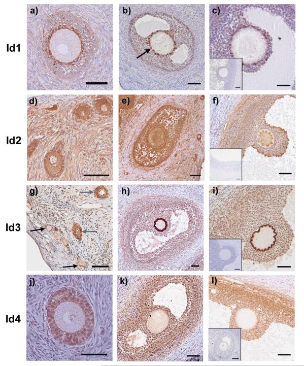
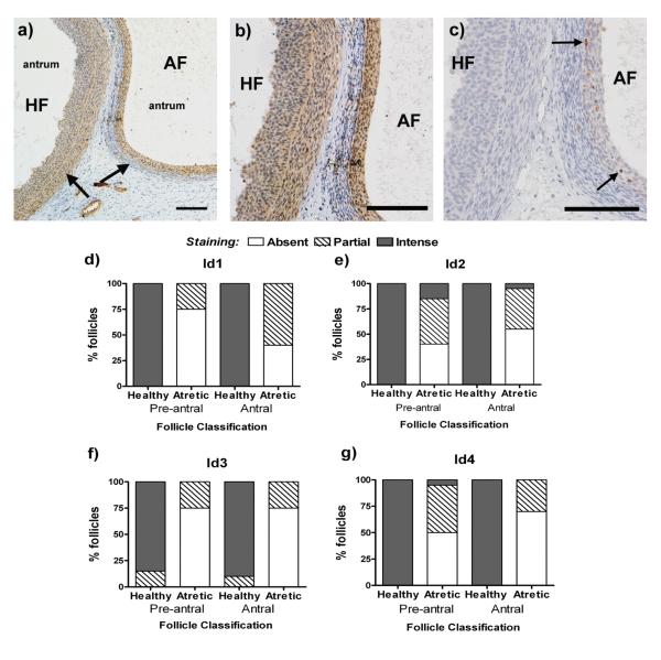
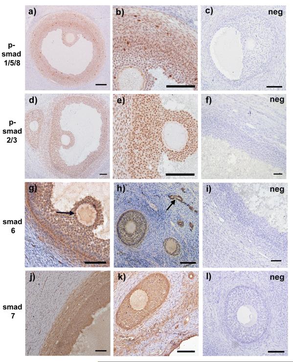
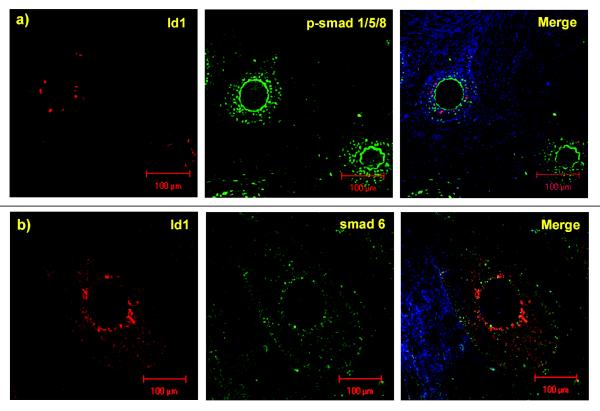
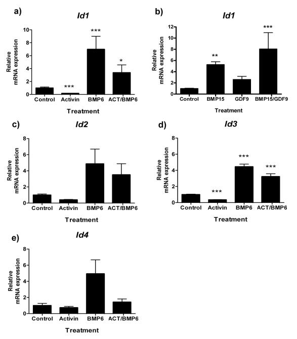
Inhibitor of differentiation (Id) proteins act during embryogenesis and development to repress gene transcription required for lineage commitment, while promoting cell growth. Growth factors belonging to the TGFbeta superfamily of signaling molecules, notably the bone morphogenetic proteins (BMPs) and activin, can regulate Id expression in these tissues. Id expression and function in adult physiology is less well determined, and we hypothesized a role for Id proteins in the adult mammalian ovary. Immunohistochemistry for Id1, Id2, Id3, and Id4 in the sheep ovary revealed consistent expression in granulosa and thecal cells of ovarian follicles throughout development. In atretic follicles, Id proteins were selectively down-regulated in thecal cells (P < 0.0001). Additionally, Id1 was universally up-regulated in the cumulus cells adjacent to the oocyte. Immunohistochemistry for phospho (p)-smad 1/5/8 signaling components (stimulated by BMPs) showed a punctate pattern of expression whereas p-smad 2/3 (stimulated by activin) was ubiquitously expressed in follicles. Neither pathway, however, displayed differential staining in line with Id1 cumulus-specific expression, suggesting a more complex relationship between Id1 expression and TGFbeta signaling in these cells. Nevertheless, in vitro, stimulation of ovine granulosa cells with BMP6 or activin A led to a respective increase and decrease in Id1 (P < 0.0001), Id2 (P < 0.0001), Id3 (P < 0.0001), and Id4 (P < 0.05) transcripts, and Id1 gene expression was further manipulated by the oocyte-secreted factors BMP15 and growth differentiation factor 9 (P < 0.001). These data confirm that TGFbeta signaling can regulate Id gene expression in the sheep ovarian follicle and suggest a functional role for the Id family in the mammalian ovary.
Figures
Similar articles
-
Characterization of Inhibitor of differentiation (Id) proteins in human cornea.Exp Eye Res. 2016 May;146:145-153. doi: 10.1016/j.exer.2015.12.003. Epub 2015 Dec 19. Exp Eye Res. 2016. PMID: 26712606 Free PMC article.
-
Hormonal regulation and differential actions of the helix-loop-helix transcriptional inhibitors of differentiation (Id1, Id2, Id3, and Id4) in Sertoli cells.Endocrinology. 2001 May;142(5):1727-36. doi: 10.1210/endo.142.5.8134. Endocrinology. 2001. PMID: 11316735
-
Expression and localization of inhibitor of differentiation (ID) proteins during tissue and vascular remodelling in the human corpus luteum.Mol Hum Reprod. 2013 Feb;19(2):82-92. doi: 10.1093/molehr/gas052. Epub 2012 Nov 16. Mol Hum Reprod. 2013. PMID: 23160862
-
Recruitment and development of the follicle; the roles of the transforming growth factor-beta superfamily.Mol Cell Endocrinol. 2002 May 31;191(1):35-43. doi: 10.1016/s0303-7207(02)00053-9. Mol Cell Endocrinol. 2002. PMID: 12044917 Review.
-
Local roles of TGF-beta superfamily members in the control of ovarian follicle development.Anim Reprod Sci. 2003 Oct 15;78(3-4):165-83. doi: 10.1016/s0378-4320(03)00089-7. Anim Reprod Sci. 2003. PMID: 12818643 Review.
Cited by
-
Characterization of Inhibitor of differentiation (Id) proteins in human cornea.Exp Eye Res. 2016 May;146:145-153. doi: 10.1016/j.exer.2015.12.003. Epub 2015 Dec 19. Exp Eye Res. 2016. PMID: 26712606 Free PMC article.
-
Inhibin removes the inhibitory effects of activin on steroid enzyme expression and androgen production by normal ovarian thecal cells.J Mol Endocrinol. 2012 Jan 25;48(1):49-60. doi: 10.1530/JME-11-0134. Print 2012 Feb. J Mol Endocrinol. 2012. PMID: 22082494 Free PMC article.
-
A high concentration of genistein down-regulates activin A, Smad3 and other TGF-β pathway genes in human uterine leiomyoma cells.Exp Mol Med. 2012 Apr 30;44(4):281-92. doi: 10.3858/emm.2012.44.4.024. Exp Mol Med. 2012. PMID: 22228119 Free PMC article.
-
Expression of genes associated with BMP signaling pathway in porcine oocytes before and after IVM - a microarray approach.Reprod Biol Endocrinol. 2017 Jun 2;15(1):43. doi: 10.1186/s12958-017-0261-6. Reprod Biol Endocrinol. 2017. PMID: 28576120 Free PMC article.
-
Functional link between bone morphogenetic proteins and insulin-like peptide 3 signaling in modulating ovarian androgen production.Proc Natl Acad Sci U S A. 2013 Apr 9;110(15):E1426-35. doi: 10.1073/pnas.1222216110. Epub 2013 Mar 25. Proc Natl Acad Sci U S A. 2013. PMID: 23530236 Free PMC article.
References
-
- Murre C, McCaw PS, Vaessin H, Caudy M, Jan LY, Jan YN, Cabrera CV, Buskin JN, Hauschka SD, Lassar AB, Weintraub H, Baltimore D. Interactions between heterologous helix-loop-helix proteins generate complexes that bind specifically to a common DNA sequence. Cell. 1989;58:537–544. - PubMed
-
- O’Toole PJ, Inoue T, Emerson L, Morrison IEG, Mackie AR, Cherry RJ, Norton JD. Id Proteins Negatively Regulate Basic Helix-Loop-Helix Transcription Factor Function by Disrupting Subnuclear Compartmentalization. J Biol Chem. 2003;278:45770–45776. - PubMed
