. 2010 Mar;84(5):2384-94.
doi: 10.1128/JVI.01686-09. Epub 2009 Dec 23.

Herpes simplex virus type 1 immediate-early protein ICP22 is required for VICE domain formation during productive viral infection

Affiliations

Herpes simplex virus type 1 immediate-early protein ICP22 is required for VICE domain formation during productive viral infection

Thomas W Bastian et al. J Virol. 2010 Mar.

Abstract

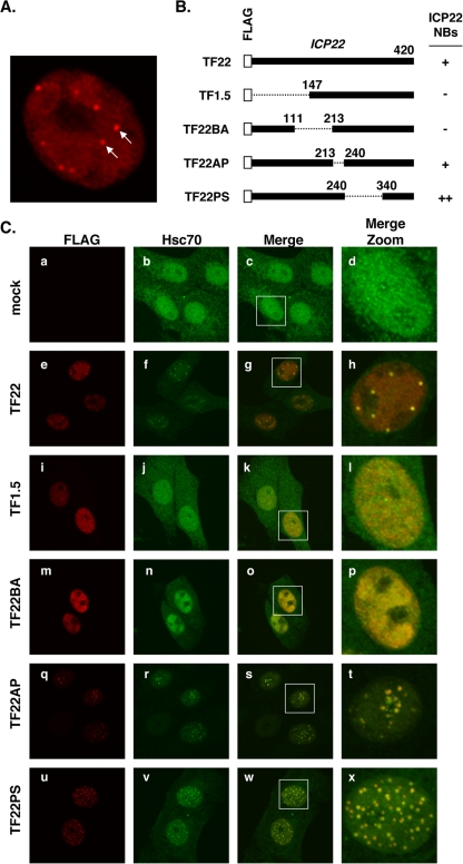
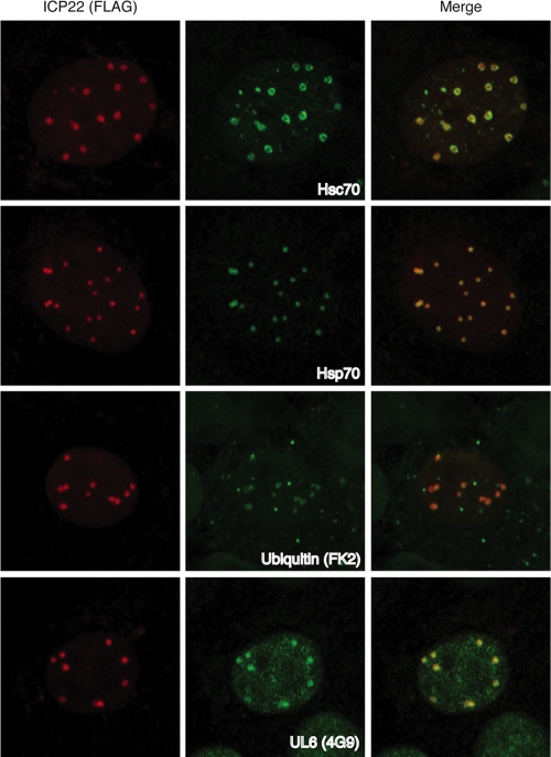
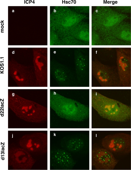
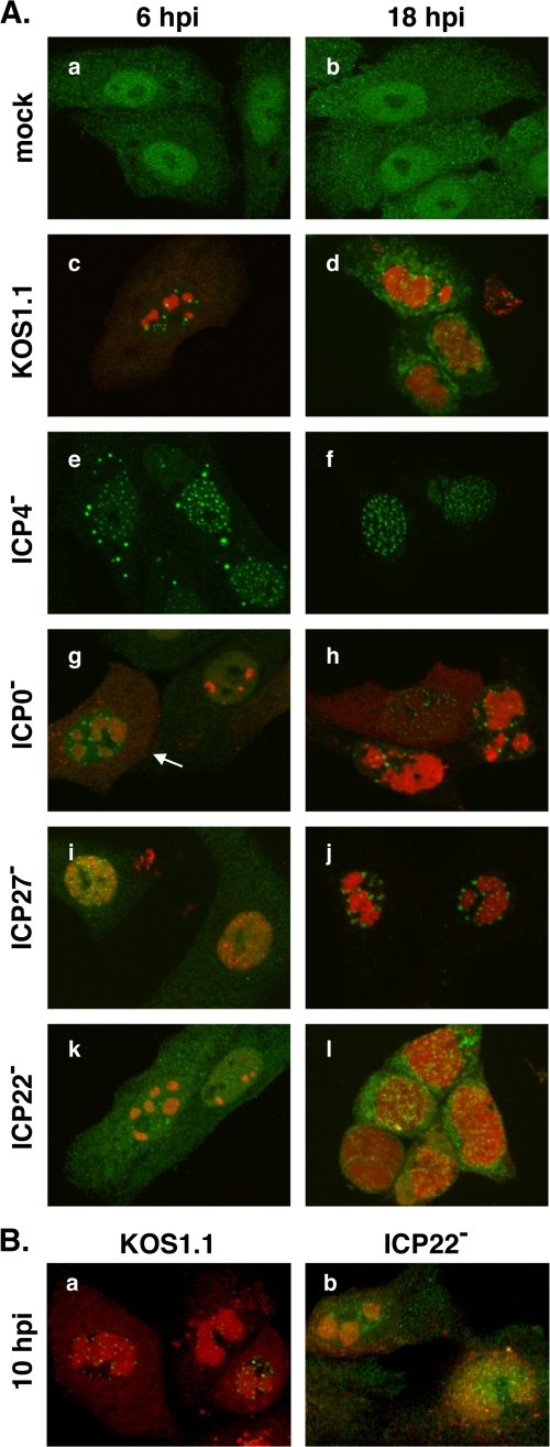
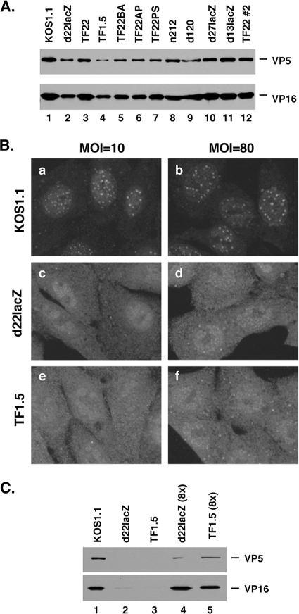
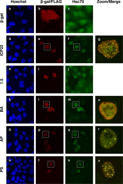
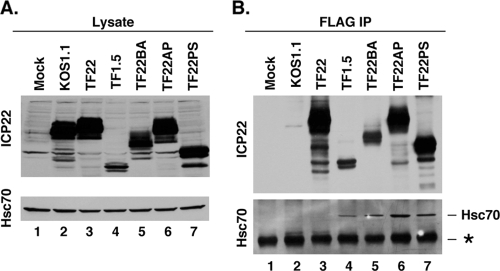
During productive infection, herpes simplex virus type 1 (HSV-1) induces the formation of discrete nuclear foci containing cellular chaperone proteins, proteasomal components, and ubiquitinated proteins. These structures are known as VICE domains and are hypothesized to play an important role in protein turnover and nuclear remodeling in HSV-1-infected cells. Here we show that VICE domain formation in Vero and other cells requires the HSV-1 immediate-early protein ICP22. Since ICP22 null mutants replicate efficiently in Vero cells despite being unable to induce VICE domain formation, it can be concluded that VICE domain formation is not essential for HSV-1 productive infection. However, our findings do not exclude the possibility that VICE domain formation is required for viral replication in cells that are nonpermissive for ICP22 mutants. Our studies also show that ICP22 itself localizes to VICE domains, suggesting that it could play a role in forming these structures. Consistent with this, we found that ICP22 expression in transfected cells is sufficient to reorganize the VICE domain component Hsc70 into nuclear inclusion bodies that resemble VICE domains. An N-terminal segment of ICP22, corresponding to residues 1 to 146, is critical for VICE domain formation in infected cells and Hsc70 reorganization in transfected cells. We previously found that this portion of the protein is dispensable for ICP22's effects on RNA polymerase II phosphorylation. Thus, ICP22 mediates two distinct regulatory activities that both modify important components of the host cell nucleus.


Figures

FIG. 1.
FIG. 1.
Colocalization of ICP22 and VICE domains. (A) Immunofluorescent staining of ICP22 in HSV-1-infected cells. Vero cells were infected at an MOI of 3 PFU/cell with HSV-1 strain TF22, encoding a FLAG-tagged ICP22. Cells were fixed at 6 hpi and stained with a FLAG-specific antiserum. The arrows point to two of the several NB structures that stained positively for ICP22. (B) Diagram of FLAG-tagged ICP22 polypeptides encoded by TF22 and related mutants. The propensity of the viruses to localize ICP22 to NBs (from reference 1) is indicated. (C) Localization of ICP22 and Hsc70 during infection. Vero cells were mock infected or infected with the indicated viruses at an MOI of 3 PFU/cell. Cells were fixed at 6 hpi and processed for immunofluorescence using antibodies specific for FLAG and Hsc70. The right-hand column shows enlargements of the images contained within the white squares.
FIG. 2.
FIG. 2.
ICP22 NBs colocalize with markers of VICE domains. Vero cells transiently expressing FLAG-tagged ICP22 were infected with strain KOS at an MOI of 10 for 6 h. Cells were colabeled with antibodies recognizing the FLAG tag and one marker of VICE domains: Hsc70, Hsp70, mono/polyubiquitinated proteins (FK2 monoclonal antibody), or the HSV-1 capsid portal protein UL6 (4G9 monoclonal antibody). Imaging was performed using a 63× 1.4 objective lens and a Zeiss LSM 510 Meta confocal microscope. Images were collected at 2× digital zoom.
FIG. 3.
FIG. 3.
ICP22 is required for formation of VICE domains. Vero cells were mock infected or infected at an MOI of 3 with WT HSV-1 or strains with mutations in ICP22 (d22lacZ) or UL13 (d13lacZ). At 6 hpi, the cells were fixed and VICE domain formation was assessed by immunofluorescence using antibodies directed against ICP4 and Hsc70.
FIG. 4.
FIG. 4.
VICE domain formation in cells infected with IE mutants. (A) Vero cells were mock infected or infected at an MOI of 3 with WT strain KOS1.1 or null mutants in ICP4 (d120), ICP0 (n212), ICP27 (d27lacZ), or ICP22 (d22lacZ). The cells were fixed at 6 and 18 hpi, and VICE domain formation was analyzed by immunofluorescence as in Fig. 3. The arrow in panel g indicates an n212-infected cell nucleus that is positive for VICE domains. (B) Vero cells were infected as for panel A, with KOS1.1 or d22lacZ. The cells were fixed at 10 hpi and analyzed by immunofluorescence as for panel A.
FIG. 5.
FIG. 5.
The inability of ICP22 mutants to induce VICE domains is not due to altered particle-to-PFU ratios. (A) Determination of relative particle-to-PFU content in various viral stocks. Aliquots of 1 × 10⁷ PFU of viral stocks, determined by plaque assay on the appropriate permissive cell line, were analyzed by SDS-PAGE and immunoblotting for the major capsid protein VP5 and tegument protein VP16. Note that two different stocks of TF22 were analyzed. (B) Vero cells were infected with the viruses shown, at an MOI of 10 or 80. At 6 hpi, cells were fixed and analyzed for VICE domains by Hsc70 staining. (C) Determination of VP5 and VP16 content in 1 × 10⁷ PFU of KOS1.1 (lane 1), d22lacZ (lane 2), or TF1.5 (lane 3) or in 8 × 10⁷ PFU of d22lacZ (lane 4) or TF1.5 (lane 5).
FIG. 6.
FIG. 6.
ICP22 can reorganize Hsc70 in transfected cells. Vero cells were transfected with expression plasmids encoding β-galactosidase, ICP22, or mutant forms of ICP22 as illustrated in Fig. 1B. One day later, the cells were fixed and processed for immunofluorescence to detect the expressed proteins and Hsc70. Note that expression of WT ICP22 as well as the BA, AP, and PS mutant forms of ICP22 increased the nuclear staining of Hsc70. For these constructs, the far right panels show merged enlargements of the images contained within the white squares.
FIG. 7.
FIG. 7.
Interaction of Hsc70 with mutant forms of ICP22. HeLa cells were mock infected or infected at an MOI of 10 with the mutants shown. At 6 hpi, total cell lysates were made and analyzed (A). (B) FLAG-tagged proteins were immunoprecipitated by incubation with affinity resin bearing anti-FLAG monoclonal antibody M2. After washing the immunoprecipitates, bound proteins were released and analyzed by immunoblotting with ICP22- or Hsc70-specific antisera. The asterisk in panel B denotes a nonspecific band corresponding to the mouse IgG heavy chain.

Similar articles

Cited by

References

    1. Bastian, T. W., and S. A. Rice. 2009. Identification of sequences in herpes simplex virus type 1 ICP22 that influence RNA polymerase II modification and viral late gene expression. J. Virol. 83:128-139. - PMC - PubMed
    1. Besse, S., and F. Puvion-Dutilleul. 1996. Intranuclear retention of ribosomal RNAs in response to herpes simplex virus type 1 infection. J. Cell Sci. 109:119-129. - PubMed
    1. Brandt, C. R., A. W. Kolb, D. D. Shah, A. M. Pumfery, R. L. Kintner, E. Jaehnig, and J. J. Van Gompel. 2003. Multiple determinants contribute to the virulence of HSV ocular and CNS infection and identification of serine 34 of the US1 gene as an ocular disease determinant. Invest. Ophthalmol. Vis. Sci. 44:2657-2668. - PubMed
    1. Bukau, B., J. Weissman, and A. Horwich. 2006. Molecular chaperones and protein quality control. Cell 125:443-451. - PubMed
    1. Burch, A. D., and S. K. Weller. 2005. Herpes simplex virus type 1 DNA polymerase requires the mammalian chaperone hsp90 for proper localization to the nucleus. J. Virol. 79:10740-10749. - PMC - PubMed

Publication types

MeSH terms

Substances