

Infection of cell lines with experimental and natural ovine scrapie agents
- PMID: 20032176
- PMCID: PMC2820909
- DOI: 10.1128/JVI.01855-09
Infection of cell lines with experimental and natural ovine scrapie agents
Abstract
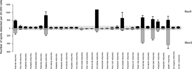
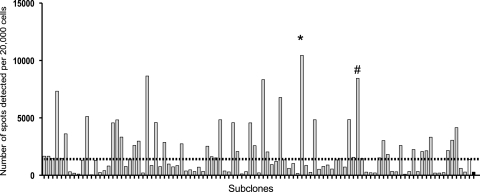
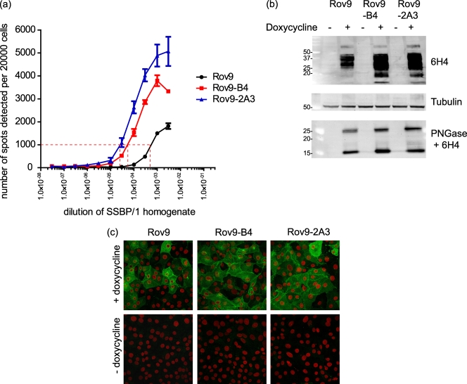
Mouse bioassay remains the gold standard for determining proof of infectivity, strain type, and infectious titer estimation in prion disease research. The development of an approach using ex vivo cell-based assays remains an attractive alternative, both in order to reduce the use of mice and to hasten results. The main limitation of a cell-based approach is the scarcity of cell lines permissive to infection with natural transmissible spongiform encephalopathy strains. This study combines two advances in this area, namely, the standard scrapie cell assay (SSCA) and the Rov9 and MovS6 cell lines, which both express the ovine PrP VRQ allele, to assess to what extent natural and experimental ovine scrapie can be detected ex vivo. Despite the Rov9 and MovS6 cell lines being of different biological origin, they were both permissive and resistant to infection with the same isolates of natural sheep scrapie as detected by SSCA. Rov9 subclones that are 20 times more sensitive than Rov9 to SSBP/1-like scrapie infection were isolated, but all the subclones maintained their resistance to isolates that failed to transmit to the parental line. The most sensitive subclone of the Rov9 cell line was used to estimate the infectious titer of a scrapie brain pool (RBP1) and proved to be more sensitive than the mouse bioassay using wild-type mice. Increasing the sensitivity of the Rov9 cell line to SSBP/1 infection did not correlate with broadening susceptibility, as the specificity of permissiveness and resistance to other scrapie isolates was maintained.
Figures
Similar articles
-
Molecular analysis of the protease-resistant prion protein in scrapie and bovine spongiform encephalopathy transmitted to ovine transgenic and wild-type mice.J Virol. 2004 Jun;78(12):6243-51. doi: 10.1128/JVI.78.12.6243-6251.2004. J Virol. 2004. PMID: 15163717 Free PMC article.
-
Sheep scrapie susceptibility-linked polymorphisms do not modulate the initial binding of cellular to disease-associated prion protein prior to conversion.J Gen Virol. 2005 Sep;86(Pt 9):2627-2634. doi: 10.1099/vir.0.80901-0. J Gen Virol. 2005. PMID: 16099922
-
A C-terminal protease-resistant prion fragment distinguishes ovine "CH1641-like" scrapie from bovine classical and L-Type BSE in ovine transgenic mice.PLoS Pathog. 2008 Aug 29;4(8):e1000137. doi: 10.1371/journal.ppat.1000137. PLoS Pathog. 2008. PMID: 18769714 Free PMC article.
-
New in vivo and ex vivo models for the experimental study of sheep scrapie: development and perspectives.C R Biol. 2002 Jan;325(1):49-57. doi: 10.1016/s1631-0691(02)01393-8. C R Biol. 2002. PMID: 11862622 Review.
-
Searching for anti-prion compounds: cell-based high-throughput in vitro assays and animal testing strategies.Methods Enzymol. 2006;412:223-34. doi: 10.1016/S0076-6879(06)12014-5. Methods Enzymol. 2006. PMID: 17046661 Review.
Cited by
-
Prion disease and the innate immune system.Viruses. 2012 Dec;4(12):3389-419. doi: 10.3390/v4123389. Viruses. 2012. PMID: 23342365 Free PMC article. Review.
-
Cellular aspects of prion replication in vitro.Viruses. 2013 Jan 22;5(1):374-405. doi: 10.3390/v5010374. Viruses. 2013. PMID: 23340381 Free PMC article. Review.
-
Enzymatic formulation capable of degrading scrapie prion under mild digestion conditions.PLoS One. 2013 Jul 16;8(7):e68099. doi: 10.1371/journal.pone.0068099. Print 2013. PLoS One. 2013. PMID: 23874511 Free PMC article.
-
Alimentary prion infections: Touchdown in the intestine.Prion. 2011 Jan-Mar;5(1):6-9. doi: 10.4161/pri.5.1.14283. Epub 2011 Jan 1. Prion. 2011. PMID: 21150306 Free PMC article.
-
Correlation of cellular factors and differential scrapie prion permissiveness in ovine microglia.Virus Res. 2017 Aug 15;240:69-80. doi: 10.1016/j.virusres.2017.07.016. Epub 2017 Jul 25. Virus Res. 2017. PMID: 28754560 Free PMC article.
References
-
- Barron, R. M., S. L. Campbell, D. King, A. Bellon, K. E. Chapman, R. A. Williamson, and J. C. Manson. 2007. High titers of transmissible spongiform encephalopathy infectivity associated with extremely low levels of PrPSc in vivo. J. Biol. Chem. 282:35878-35886. - PubMed
-
- Baylis, M., C. Chihota, E. Stevenson, W. Goldmann, A. Smith, K. Sivam, S. Tongue, and M. B. Gravenor. 2004. Risk of scrapie in British sheep of different prion protein genotype. J. Gen. Virol. 85:2735-2740. - PubMed
-
- Brightwell, G., V. Poirier, E. Cole, S. Ivins, and K. W. Brown. 1997. Serum-dependent and cell cycle-dependent expression from a cytomegalovirus-based mammalian expression vector. Gene 194:115-123. - PubMed
Publication types
MeSH terms
Substances
LinkOut - more resources
Full Text Sources
Research Materials
Miscellaneous
