Contributions of the avian influenza virus HA, NA, and M2 surface proteins to the induction of neutralizing antibodies and protective immunity
- PMID: 20032181
- PMCID: PMC2820928
- DOI: 10.1128/JVI.02135-09
Contributions of the avian influenza virus HA, NA, and M2 surface proteins to the induction of neutralizing antibodies and protective immunity
Abstract
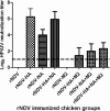
Highly pathogenic avian influenza virus (HPAIV) subtype H5N1 causes severe disease and mortality in poultry. Increased transmission of H5N1 HPAIV from birds to humans is a serious threat to public health. We evaluated the individual contributions of each of the three HPAIV surface proteins, namely, the hemagglutinin (HA), the neuraminidase (NA), and the M2 proteins, to the induction of HPAIV-neutralizing serum antibodies and protective immunity in chickens. Using reverse genetics, three recombinant Newcastle disease viruses (rNDVs) were engineered, each expressing the HA, NA, or M2 protein of H5N1 HPAIV. Chickens were immunized with NDVs expressing a single antigen (HA, NA, and M2), two antigens (HA+NA, HA+M2, and NA+M2), or three antigens (HA+NA+M2). Immunization with HA or NA induced high titers of HPAIV-neutralizing serum antibodies, with the response to HA being greater, thus identifying HA and NA as independent neutralization antigens. M2 did not induce a detectable neutralizing serum antibody response, and inclusion of M2 with HA or NA reduced the magnitude of the response. Immunization with HA alone or in combination with NA induced complete protection against HPAIV challenge. Immunization with NA alone or in combination with M2 did not prevent death following challenge, but extended the time period before death. Immunization with M2 alone had no effect on morbidity or mortality. Thus, there was no indication that M2 is immunogenic or protective. Furthermore, inclusion of NA in addition to HA in a vaccine preparation for chickens may not enhance the high level of protection provided by HA.
Figures


Similar articles
-
Coexpression of avian influenza virus H5 and N1 by recombinant Newcastle disease virus and the impact on immune response in chickens.Avian Dis. 2011 Sep;55(3):413-21. doi: 10.1637/9652-011111-Reg.1. Avian Dis. 2011. PMID: 22017039
-
Newcastle disease virus-vectored vaccines expressing the hemagglutinin or neuraminidase protein of H5N1 highly pathogenic avian influenza virus protect against virus challenge in monkeys.J Virol. 2010 Feb;84(3):1489-503. doi: 10.1128/JVI.01946-09. Epub 2009 Nov 18. J Virol. 2010. PMID: 19923177 Free PMC article.
-
A recombinant avian paramyxovirus serotype 3 expressing the hemagglutinin protein protects chickens against H5N1 highly pathogenic avian influenza virus challenge.Sci Rep. 2020 Feb 10;10(1):2221. doi: 10.1038/s41598-020-59124-x. Sci Rep. 2020. PMID: 32042001 Free PMC article.
-
Contribution of antibody production against neuraminidase to the protection afforded by influenza vaccines.Rev Med Virol. 2012 Jul;22(4):267-79. doi: 10.1002/rmv.1713. Epub 2012 Mar 22. Rev Med Virol. 2012. PMID: 22438243 Free PMC article. Review.
-
In the shadow of hemagglutinin: a growing interest in influenza viral neuraminidase and its role as a vaccine antigen.Viruses. 2014 Jun 23;6(6):2465-94. doi: 10.3390/v6062465. Viruses. 2014. PMID: 24960271 Free PMC article. Review.
Cited by
-
Evaluation of the Newcastle disease virus F and HN proteins in protective immunity by using a recombinant avian paramyxovirus type 3 vector in chickens.J Virol. 2011 Jul;85(13):6521-34. doi: 10.1128/JVI.00367-11. Epub 2011 Apr 27. J Virol. 2011. Retraction in: J Virol. 2020 Feb 28;94(6):e01867-19. doi: 10.1128/JVI.01867-19. PMID: 21525340 Free PMC article. Retracted.
-
N-linked glycosylation in the hemagglutinin of influenza A viruses.Yonsei Med J. 2012 Sep;53(5):886-93. doi: 10.3349/ymj.2012.53.5.886. Yonsei Med J. 2012. PMID: 22869469 Free PMC article. Review.
-
Evaluation of the replication, pathogenicity, and immunogenicity of avian paramyxovirus (APMV) serotypes 2, 3, 4, 5, 7, and 9 in rhesus macaques.PLoS One. 2013 Oct 10;8(10):e75456. doi: 10.1371/journal.pone.0075456. eCollection 2013. PLoS One. 2013. Retraction in: PLoS One. 2020 Dec 14;15(12):e0244045. doi: 10.1371/journal.pone.0244045. PMID: 24130713 Free PMC article. Retracted.
-
H5Nx Viruses Emerged during the Suppression of H5N1 Virus Populations in Poultry.Microbiol Spectr. 2021 Oct 31;9(2):e0130921. doi: 10.1128/Spectrum.01309-21. Epub 2021 Sep 29. Microbiol Spectr. 2021. PMID: 34585974 Free PMC article.
-
Influenza Neuraminidase Characteristics and Potential as a Vaccine Target.Front Immunol. 2021 Nov 16;12:786617. doi: 10.3389/fimmu.2021.786617. eCollection 2021. Front Immunol. 2021. PMID: 34868073 Free PMC article. Review.
References
-
- Alexander, D. J. 2007. An overview of the epidemiology of avian influenza. Vaccine 25:5637-5644. - PubMed
-
- Chen, Q., H. Kuang, H. Wang, F. Fang, Z. Yang, Z. Zhang, X. Zhang, and Z. Chen. 2009. Comparing the ability of a series of viral protein-expressing plasmid DNAs to protect against H5N1 influenza virus. Virus Genes 38:30-38. - PubMed
-
- Chen, Z., Y. Sahashi, K. Matsuo, H. Asanuma, H. Takahashi, T. Iwasaki, Y. Suzuki, C. Aizawa, T. Kurata, and S. Tamura. 1998. Comparison of the ability of viral protein-expressing plasmid DNAs to protect against influenza. Vaccine 16:1544-1549. - PubMed
-
- Cleverley, D. Z., H. M. Geller, and J. Lenard. 1997. Characterization of cholesterol-free insect cells infectible by baculoviruses: effects of cholesterol on VSV fusion and infectivity and on cytotoxicity induced by influenza M2 protein. Exp. Cell Res. 233:288-296. - PubMed
-
- Denis, J., E. Acosta-Ramirez, Y. Zhao, M. E. Hamelin, I. Koukavica, M. Baz, Y. Abed, C. Savard, C. Pare, C. Lopez Macias, G. Boivin, and D. Leclerc. 2008. Development of a universal influenza A vaccine based on the M2e peptide fused to the papaya mosaic virus (PapMV) vaccine platform. Vaccine 26:3395-3403. - PubMed
Publication types
MeSH terms
Substances
Grants and funding
LinkOut - more resources
Full Text Sources
Other Literature Sources
Medical

