. 2010 Mar;84(6):2832-42.
doi: 10.1128/JVI.02435-09. Epub 2009 Dec 23.

The foamy virus genome remains unintegrated in the nuclei of G1/S phase-arrested cells, and integrase is critical for preintegration complex transport into the nucleus

Affiliations

The foamy virus genome remains unintegrated in the nuclei of G1/S phase-arrested cells, and integrase is critical for preintegration complex transport into the nucleus

Yung-Tsun Lo et al. J Virol. 2010 Mar.

Abstract

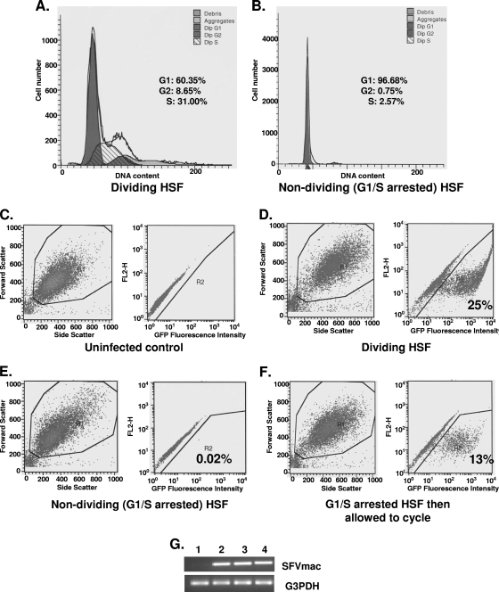
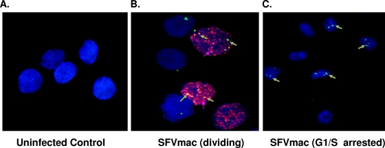
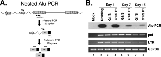
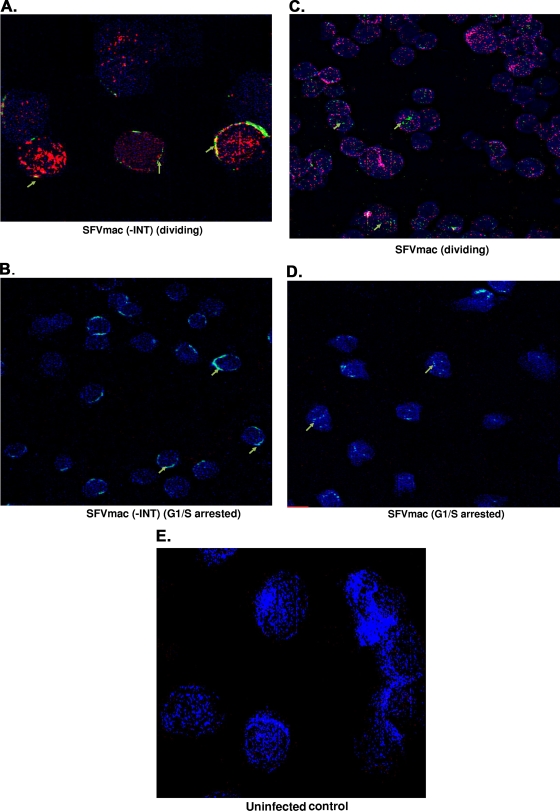
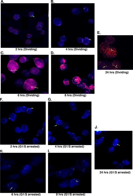
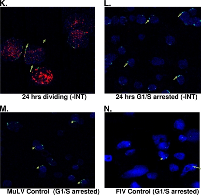
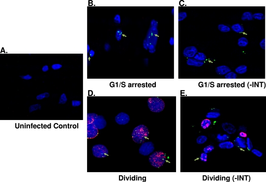
Foamy viruses are a member of the spumavirus subfamily of retroviruses with unique mechanisms of virus replication. Foamy virus replication is cell cycle dependent; however, the genome is found in the nuclei of cells arrested in the G(1)/S phase. Despite the presence of genome in the nuclei of growth-arrested cells, there is no viral gene expression, thus explaining its dependency on cell cycle. This report shows that the foamy virus genome remains unintegrated in G(1)/S phase-arrested cells. The foamy virus genome is detected by confocal microscopy in the nuclei of both dividing and growth-arrested cells. Alu PCR revealed foamy virus-specific DNA amplification from genomic DNA isolated in cycling cells at 24 h postinfection. In arrested cells no foamy virus DNA band was detected in cells harvested at 1 or 7 days after infection, and a very faint band that is significantly less than DNA amplified from cycling cells was observed at day 15. After these cells were arrested at the G(1)/S phase for 1, 7, or 15 days they were allowed to cycle, at which time foamy virus-specific DNA amplification was readily observed. Taken together, these results suggest that the foamy virus genome persists in nondividing cells without integrating. We have also established evidence for the first time that the foamy virus genome and Gag translocation into the nucleus are dependent on integrase in cycling cells, implicating the role of integrase in transport of the preintegration complex into the nucleus. Furthermore, despite the presence of a nuclear localization signal sequence in Gag, we observed no foamy virus Gag importation into the nucleus in the absence of integrase.


Figures

FIG. 1.
FIG. 1.
Transduction and transgene expression efficiency by SFVmac in cycling and G1/S phase-arrested HSF cells. Cell cycle analysis results in cycling and arrested cells are shown in panels A and B, respectively. The populations of cells at different phases of the cell cycle are given as a percentage of the total population. Cells in G1/S phase were at a 97% level. FACS analyses were performed at the same time for GFP expression. (C) FACS results for GFP expression in uninfected dividing cells. (D and E) FACS results of dividing and growth-arrested GFP-expressing cells, respectively, infected with SFVmac vector. (F) GFP expression in cells where medium containing aphidicolin was removed from the culture and replaced with fresh medium without aphidicolin, allowing cells to undergo cell division. (G) DNA amplified by PCR from SFVmac vector-transduced HSF cells. Lane 1 is mock transduced cells, lane 2 is cycling HSF cells transduced with SFVmac vector, and lane 3 represents G1/S phase-arrested cells transduced with foamy vector. PCR results from growth-arrested cells infected with foamy virus vector and released into cycle 24 h later are shown in lane 4. DNA was amplified using pol-specific forward 5′-TGTAATACCACTCCAAGCCTGGAT-3′ and reverse 5′-GACTTTCAGAAAAGTAGCGTCTCG-3′ primers.
FIG. 2.
FIG. 2.
SFVmac genome localization in dividing and growth-arrested cells. For confocal microscopy cells were labeled with BrdU (red) to distinguish between dividing and growth-arrested cells. SFV-1 genome was identified with in situ hybridization using gag-specific probe (green). The nucleus was counterstained with DAPI (blue). (A) In situ staining of uninfected control cells. (B and C) Dividing and growth-arrested cells, respectively, transduced with foamy virus vector.
FIG. 3.
FIG. 3.
Alu PCR of genomic DNA isolated from SFVmac vector-infected dividing or growth-arrested cells. (A) Schematic representation of an Alu PCR methodology. DNA was copied using an Alu sequence-specific primer and amplified with an SFVmac LTR-specific primer tagged with a lambda sequence. (B) Alu PCR with samples isolated from dividing and growth-arrested cells (top panel). The Alu PCR-amplified products were hybridized to radioactive [γ-32P]ATP-labeled LTR-specific probe. Lane 1 (mock) contains PCR results from uninfected control cells. Lane 2 (dividing) is the PCR product with DNA isolated from SFVmac-transduced dividing cells 24 h after transduction. Lanes 3, 5, and 7 represent PCR results from G1/S-arrested cells harvested at days 1, 7, and 15 posttransduction, respectively. Lanes 4, 6, and 8 are PCR products for cells that were arrested in G1/S for 1, 7, and 15 days, respectively, and then allowed to resume a normal cycle for 72 h. The second and third panels represent corresponding semiquantitative regular PCR detecting the pol and LTR region of SFVmac. For level of sample recovery, the DNAs were amplified with primers specific to G3PDH (glyceraldehyde-3-phosphate dehydrogenase) (bottom panel).
FIG. 4.
FIG. 4.
SFVmac genome localization in the absence of integrase. Cycling cells or growth-arrested cells were transduced with SFVmac vector prepared in the absence of integrase. Permeabilized cells were labeled with anti-Nup153 for nucleoporin 153 of the nuclear membrane (blue). Cells were also labeled with BrdU to identify dividing cells (red), and in situ hybridization was performed to label SFVmac genome (green). (A) Dividing cells transduced with SFVmac vector with an integrase deficient SFVmac vector. (B) Nondividing cells transduced with the same SFVmac vector. (C and D) Images representative of dividing and growth-arrested cells, respectively, transduced with an SFVmac vector with intact integrase. An uninfected control is shown in panel E. “-INT” indicates integrase-defective SFVmac vector.
FIG. 5.
FIG. 5.
SFVmac genome localization in the nuclei of nucleoporin labeled dividing and growth-arrested cells at different time points after infection with SFVmac vector. BrdU (red)-labeled cells were permeabilized, and in situ hybridization was performed by probing for SFVmac genome (green). The nucleus was defined by immunostaining with antibody to nucleoporin, Nup153 (blue). (A to E) In situ hybridization for foamy virus genome with immunolabeling for Nup153 at the indicated time points in dividing cells. (F to J) Genome localization at different time points in cells arrested at the G1/S phase. Foamy virus genome localization in dividing and growth-arrested cells infected with integrase defective vector at 24 h posttransduction is shown in panels K and L, respectively. In situ hybridization for MuLV and FIV controls at 24 h posttransduction are represented in panels M and N, respectively.
FIG. 5.
FIG. 5.
SFVmac genome localization in the nuclei of nucleoporin labeled dividing and growth-arrested cells at different time points after infection with SFVmac vector. BrdU (red)-labeled cells were permeabilized, and in situ hybridization was performed by probing for SFVmac genome (green). The nucleus was defined by immunostaining with antibody to nucleoporin, Nup153 (blue). (A to E) In situ hybridization for foamy virus genome with immunolabeling for Nup153 at the indicated time points in dividing cells. (F to J) Genome localization at different time points in cells arrested at the G1/S phase. Foamy virus genome localization in dividing and growth-arrested cells infected with integrase defective vector at 24 h posttransduction is shown in panels K and L, respectively. In situ hybridization for MuLV and FIV controls at 24 h posttransduction are represented in panels M and N, respectively.
FIG. 6.
FIG. 6.
SFV Gag localization in the absence of integrase. Dividing and nondividing cells were transduced with SFVmac vector defective for integrase, and immunohistochemistry was performed with antibody against Gag (green) and nucleoporin with anti-Nup153 (blue). BrdU labeling is represented by a red stain showing dividing cells. (A) Uninfected control. (B and C) Growth-arrested cells transduced with wild-type SFVmac vector and integrase defective vector, respectively. Gag localization in dividing cells are represented in panels D and E. “-INT” indicates integrase-defective SFVmac vector.

Similar articles

Cited by

References

    1. An, D. G., U. Hyun, and C. G. Shin. 2008. Characterization of nuclear localization signals of the prototype foamy virus integrase. J. Gen. Virol. 89:1680-1684. - PubMed
    1. Bahner, I., K. Kearns, Q. L. Hao, E. M. Smogorzewska, and D. B. Kohn. 1996. Transduction of human CD34+ hematopoietic progenitor cells by a retroviral vector expressing an RRE decoy inhibits human immunodeficiency virus type 1 replication in myelomonocytic cells produced in long-term culture. J. Virol. 70:4352-4360. - PMC - PubMed
    1. Bauer, T. R. J., J. M. Allen, M. Hai, L. M. Tuschong, I. F. Khan, E. M. Olson, R. L. Adler, T. H. Burkholder, Y. C. Gu, D. W. Russell, and D. D. Hickstein. 2008. Successful treatment of canine leukocyte adhesion deficiency by foamy virus vectors. Nat. Med. 14:93-97. - PMC - PubMed
    1. Bukovsky, A., and H. Gottlinger. 1996. Lack of integrase can markedly affect human immunodeficiency virus type 1 particle production in the presence of an active viral protease. J. Virol. 70:6820-6825. - PMC - PubMed
    1. Bukrinsky, M. I., S. Haggerty, M. P. Dempsey, N. Sharova, A. Adzhubel, L. Spitz, P. Lewis, D. Goldfarb, M. Emerman, and M. Stevenson. 1993. A nuclear localization signal within HIV-1 matrix protein that governs infection of non-dividing cells. Nature 365:666-669. - PMC - PubMed

Publication types

MeSH terms

LinkOut - more resources