. 2010 Mar;84(5):2421-31.
doi: 10.1128/JVI.02174-09. Epub 2009 Dec 23.

The interferon stimulator mitochondrial antiviral signaling protein facilitates cell death by disrupting the mitochondrial membrane potential and by activating caspases

Affiliations

The interferon stimulator mitochondrial antiviral signaling protein facilitates cell death by disrupting the mitochondrial membrane potential and by activating caspases

Chia-Yi Yu et al. J Virol. 2010 Mar.

Abstract

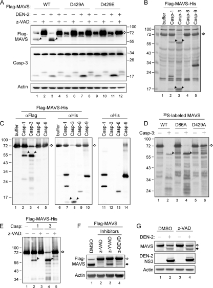
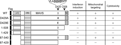
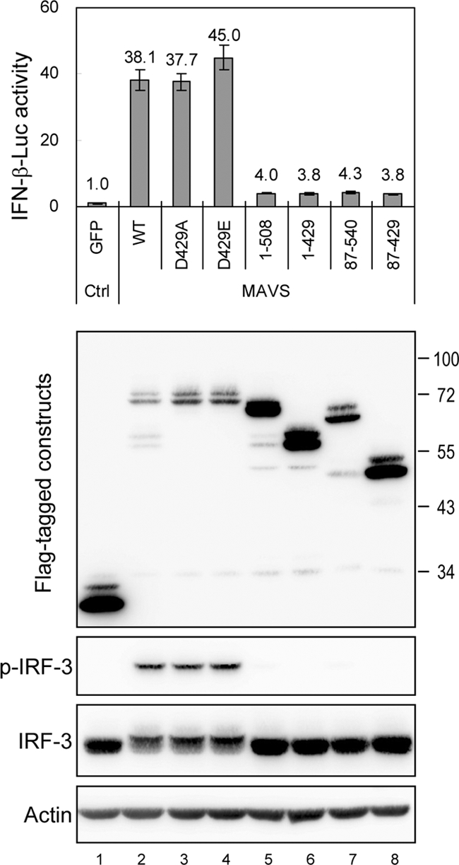
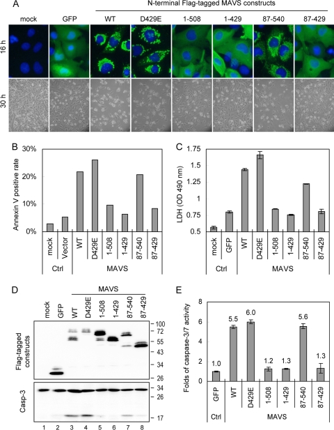
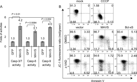
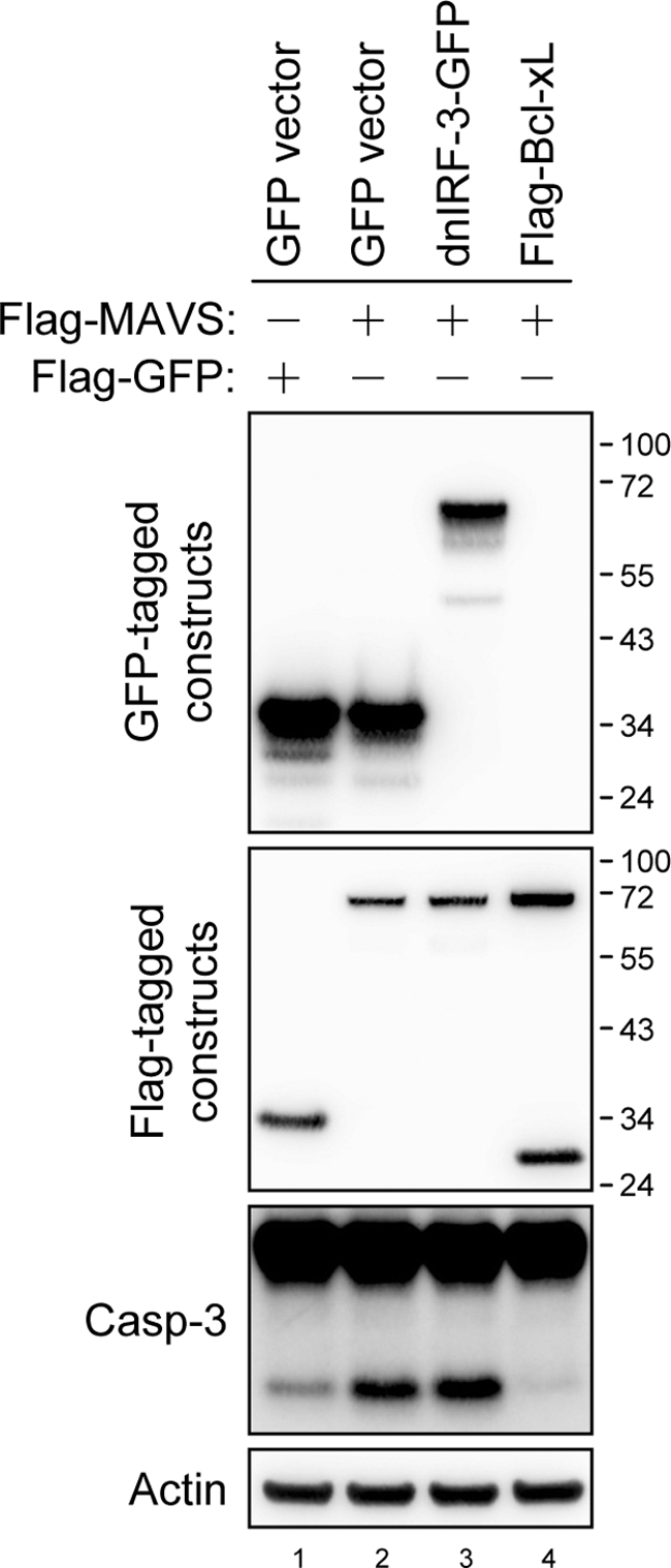
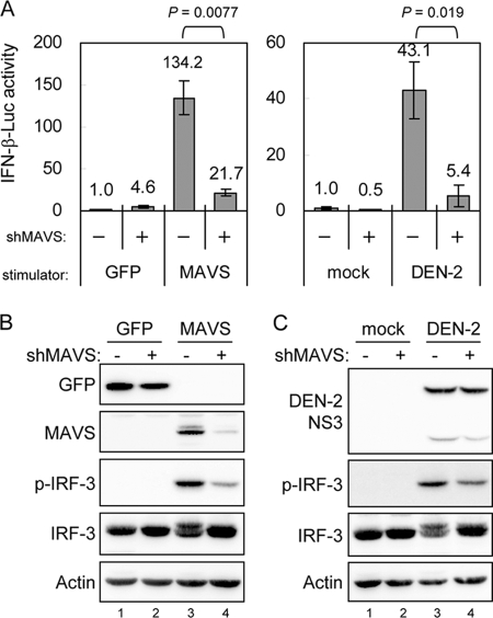
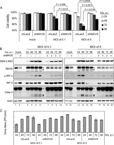
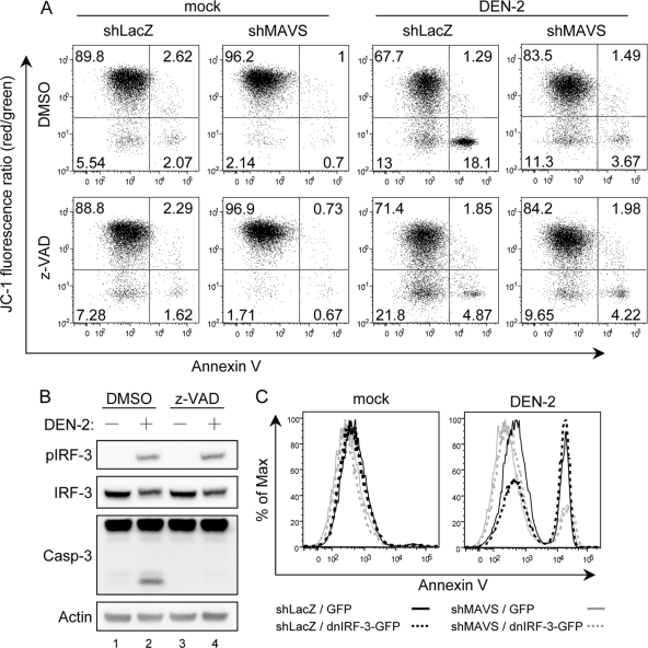
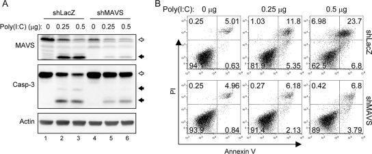
Interferon (IFN) signaling is initiated by the recognition of viral components by host pattern recognition receptors. Dengue virus (DEN) triggers IFN-beta induction through a molecular mechanism involving the cellular RIG-I/MAVS signaling pathway. Here we report that the MAVS protein level is reduced in DEN-infected cells and that caspase-1 and caspase-3 cleave MAVS at residue D429. In addition to its well-known function in IFN induction, MAVS is also a proapoptotic molecule that triggers disruption of the mitochondrial membrane potential and activation of caspases. Although different domains are required for the induction of cytotoxicity and IFN, caspase cleavage at residue 429 abolished both functions of MAVS. The apoptotic role of MAVS in viral infection and double-stranded RNA (dsRNA) stimulation was demonstrated in cells with reduced endogenous MAVS expression induced by RNA interference. Even though IFN-beta promoter activation was largely suppressed, DEN production was not affected greatly in MAVS knockdown cells. Instead, DEN- and dsRNA-induced cell death and caspase activation were delayed and attenuated in the cells with reduced levels of MAVS. These results reveal a new role of MAVS in the regulation of cell death beyond its well-known function of IFN induction in antiviral innate immunity.


Figures

FIG. 1.
FIG. 1.
Caspase-1 and caspase-3 cleave MAVS at D429. (A) N18 cells were mock treated or adsorbed with DEN-2 (multiplicity of infection [MOI] of 5) and transfected with 2 μg of wild-type (WT) or D429A- or D429E-mutated Flag-MAVS. The cells were then incubated in the presence (+) or absence (−) of z-VAD-fmk (150 μM) for 30 h. Cell lysates were analyzed by immunoblotting, using the antibodies indicated at the left. (B and C) Recombinant Flag-MAVS-His protein (30 μg) was incubated with 2 U of active caspase-1, -3, -8, or -9 as described in Materials and Methods. The reaction mixtures were resolved by SDS-PAGE and analyzed by silver staining (B) and immunoblotting, using antibodies against Flag (lanes 1 to 5) and His (lanes 6 to 10), as indicated at the top (C). The background reactivity of these caspases with anti-His antibody was also demonstrated by immunoblotting in the absence of Flag-MAVS-His protein (lanes 11 to 14 in panel C). (D) The cleavage assay of in vitro-transcribed and -translated 35S-labeled MAVS and its mutants (D86A and D429A) was performed by coincubation with (+) or without (−) active caspase-3. The proteins were then separated by SDS-PAGE, and the signals were developed by autoradiography. (E) In vitro cleavage of Flag-MAVS-His protein by caspase-1 and caspase-3 was performed in the presence (+) or absence (−) of z-VAD-fmk (1 mM) and analyzed by immunoblotting using anti-Flag antibody. (F) N18 cells were transfected with Flag-tagged MAVS and treated with various caspase inhibitors, as indicated at the top, for 24 h. Cell lysates were harvested and analyzed by immunoblotting, using anti-Flag and anti-actin antibodies, as indicated at the left. (G) A549 cells were infected with DEN-2 (MOI of 5) and then treated with z-VAD-fmk (100 μM). Cell lysates were harvested 48 h after infection and analyzed by immunoblotting, using antibodies against endogenous MAVS, DEN-2 NS3, and actin, as indicated at the left. The open arrows indicate full-length MAVS, and the black arrows indicate cleaved MAVS. DMSO, dimethyl sulfoxide.
FIG. 2.
FIG. 2.
Schematic diagram and summarized properties of MAVS constructs. The constructs were N-terminally Flag tagged and are numbered according to amino acid residues. The regions corresponding to the three domains of MAVS are also indicated, as follows: CARD, caspase recruitment domain; PR, proline-rich domain; and TM, transmembrane domain. The arrows indicate the HCV protease (C508) and caspase (D429) cleavage sites of MAVS. The abilities of IFN promoter activation, mitochondrial localization, and cytotoxicity induction of these MAVS constructs are also summarized.
FIG. 3.
FIG. 3.
Caspase-cleaved MAVS loses the ability to activate the IFN promoter. N18 cells were cotransfected with p125-Luc (0.3 μg), IRF-3/pCR3.1 (0.3 μg), pRL-TK (0.1 μg), and the indicated MAVS constructs (0.6 μg) for 24 h, and the cell lysates were harvested and analyzed by dual-luciferase assay and by Western blotting, using antibodies against Flag, Ser396-phosphorylated IRF-3, IRF-3, and actin, as indicated at the left. Firefly luciferase activity was normalized to that of Renilla luciferase, and the change in induction relative to that of the green fluorescent protein (GFP) control was determined. The results are expressed as averages and standard deviations (n = 3 per group).
FIG. 4.
FIG. 4.
Caspase-cleaved MAVS loses the ability to trigger cell death. (A) (Top) The expression pattern of each MAVS construct in N18 cells was analyzed using an immunofluorescence assay 16 h after transfection. Green, staining with anti-Flag antibody plus Alexa Fluor 488-labeled secondary antibody; blue, nuclear 4′,6-diamidino-2-phenylindole [DAPI] staining. (Bottom) Cell morphology was also photographed by phase-contrast microscopy 30 h after transfection. (B) Twenty-four hours after transfection, the transfected cells were collected and analyzed for annexin V positivity by flow cytometry. (C) The culture supernatants were collected 30 h after transfection, and cytotoxicity was estimated by measuring LDH release. The results are expressed as averages and standard deviations (n = 2 per group). (D) MAVS protein expression and caspase-3 activation were determined in N18 transfectants by immunoblotting with antibodies against the Flag tag and caspase-3, as indicated at the left. Flag-tagged GFP was used as a protein expression control. (E) The levels of caspase-3/7 activation triggered by various MAVS constructs were determined in 293FT cells transfected with the GFP control or the indicated MAVS constructs for 48 h, using Caspase-Glo 3/7 assays. The change in induction relative to that of the GFP control was determined, and the results are expressed as averages and standard deviations (n = 3 per group).
FIG. 5.
FIG. 5.
Overexpression of MAVS triggers disruption of the mitochondrial membrane potential and caspase activation. (A) The activation of caspase-3/7, -8, and -9 by MAVS overexpression was determined in transfected 293FT cells by using Caspase-Glo 3/7, 8, and 9 assays as described in the legend to Fig. 4E (n = 3 per group). (B) N18 cells were transfected with MAVS, Bcl-xS, or vector control in the presence or absence of z-VAD-fmk (100 μM) for 24 h and then analyzed by flow cytometry, using JC-1 and annexin V double staining. A decreased red/green fluorescence ratio of JC-1 represents MMP disruption, as shown in the cells treated with CCCP (50 μM), an MMP disrupter.
FIG. 6.
FIG. 6.
Bcl-xL but not dnIRF-3 blocks MAVS-induced caspase-3 activation. N18 cells were transfected with plasmids encoding dominant-negative IRF-3 fused with GFP, Flag-tagged Bcl-xL, GFP control (1 μg) plus Flag-MAVS, or Flag-GFP control (1 μg), as indicated at the top, for 24 h. Cell lysates were harvested for Western blot analyses with antibodies against GFP, the Flag tag, caspase-3, and actin, as indicated at the left.
FIG. 7.
FIG. 7.
MAVS- and virus-induced IFN signaling is attenuated in MAVS knockdown cells. (A) Lentivirus-transduced A549 cells with (+) or without (−) MAVS-targeting shRNA were transfected with p125-Luc, IRF-3/pCR3.1, pRL-TK plus GFP, or MAVS as described in the legend to Fig. 3. For DEN-2-induced IFN-β promoter analysis, the cells were adsorbed with DEN-2 (MOI = 5) for 2 h and then transfected with p125-Luc (0.6 μg) and pRL-TK (0.15 μg) for 24 h. The change in IFN-β promoter activation was determined as described in the legend to Fig. 3. The IFN-β induction of the indicated groups was compared by two-tailed Student's t test. (B and C) Cells were prepared as described for panel A, and the cell lysates were analyzed by Western blotting with the antibodies indicated at the left.
FIG. 8.
FIG. 8.
DEN-2-induced cytotoxicity is attenuated by reducing endogenous MAVS protein expression. A549-shMAVS or control (shLacZ) cells were infected with DEN-2 at an MOI of 0.1 or 5. The cells and the culture supernatants were collected at the indicated times postinfection (p.i.). (A) Cells were counted for viability counts by trypan blue exclusion. The results are expressed as averages and standard deviations (n = 3). The cell viabilities of the indicated groups were compared by two-tailed Student's t test. (B) The cells were lysed for immunoblotting using the antibodies indicated at the left. The open arrows indicate full-length MAVS or procaspase-3, and the black arrows indicate cleaved MAVS or caspase-3. (C) The supernatants were used to measure virus titers by plaque-forming assays, and the results are expressed as averages and standard deviations (n = 3).
FIG. 9.
FIG. 9.
Knockdown of MAVS attenuates DEN-2-triggered MMP disruption. (A) Mock- or DEN-2-infected (MOI of 5) cells were cultured in the presence or absence of z-VAD-fmk (100 μM) for 48 h and then analyzed by flow cytometry for JC-1 and annexin V double staining, as described in the legend to Fig. 5B. (B) The cell lysates described for Fig. 1G were analyzed with the antibodies indicated at the left. (C) Control (shLacZ) and MAVS knockdown (shMAVS) cells were mock infected or infected with DEN-2 (MOI of 5) and transfected with dnIRF-3-GFP or control GFP vector. Cells were harvested at 48 h p.i. and stained with Cy5-conjugated annexin V. GFP-positive cells were gated and analyzed for annexin V positivity by flow cytometry.
FIG. 10.
FIG. 10.
MAVS facilitates cytosolic poly(I:C)-triggered cell death. (A) The synthetic dsRNA analog poly(I:C) was delivered to A549-shMAVS or A549-shLacZ cells at the doses indicated at the top, and the cells were harvested 24 h after transfection. Endogenous MAVS protein expression and caspase-3 activation were determined by immunoblotting with antibodies against MAVS and caspase-3, respectively. The open arrows indicate full-length MAVS or procaspase-3, and the black arrows indicate cleaved MAVS or caspase-3. Immunoblotting with anti-actin is shown as a protein loading control. (B) The transfected cells were also collected for cell death analysis by flow cytometry of annexin V and PI double staining.

Similar articles

Cited by

References

    1. Andrejeva, J., K. S. Childs, D. F. Young, T. S. Carlos, N. Stock, S. Goodbourn, and R. E. Randall. 2004. The V proteins of paramyxoviruses bind the IFN-inducible RNA helicase, mda-5, and inhibit its activation of the IFN-beta promoter. Proc. Natl. Acad. Sci. USA 101:17264-17269. - PMC - PubMed
    1. Backes, C., J. Kuentzer, H. P. Lenhof, N. Comtesse, and E. Meese. 2005. GraBCas: a bioinformatics tool for score-based prediction of caspase- and granzyme B-cleavage sites in protein sequences. Nucleic Acids Res. 33:W208-W213. - PMC - PubMed
    1. Besch, R., H. Poeck, T. Hohenauer, D. Senft, G. Hacker, C. Berking, V. Hornung, S. Endres, T. Ruzicka, S. Rothenfusser, and G. Hartmann. 2009. Proapoptotic signaling induced by RIG-I and MDA-5 results in type I interferon-independent apoptosis in human melanoma cells. J. Clin. Invest. 119:2399-2411. - PMC - PubMed
    1. Bhoj, V. G., Q. Sun, E. J. Bhoj, C. Somers, X. Chen, J. P. Torres, A. Mejias, A. M. Gomez, H. Jafri, O. Ramilo, and Z. J. Chen. 2008. MAVS and MyD88 are essential for innate immunity but not cytotoxic T lymphocyte response against respiratory syncytial virus. Proc. Natl. Acad. Sci. USA 105:14046-14051. - PMC - PubMed
    1. Chang, T. H., C. L. Liao, and Y. L. Lin. 2006. Flavivirus induces interferon-beta gene expression through a pathway involving RIG-I-dependent IRF-3 and PI3K-dependent NF-kappaB activation. Microbes Infect. 8:157-171. - PubMed

Publication types

MeSH terms