. 2010 Feb;24(2):391-401.
doi: 10.1210/me.2009-0319. Epub 2009 Dec 23.

Regulation of signal transducer and activator of transcription 3 enhanceosome formation by apurinic/apyrimidinic endonuclease 1 in hepatic acute phase response

Affiliations

Regulation of signal transducer and activator of transcription 3 enhanceosome formation by apurinic/apyrimidinic endonuclease 1 in hepatic acute phase response

Sutapa Ray et al. Mol Endocrinol. 2010 Feb.

Abstract

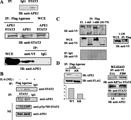
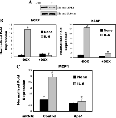
The signal transducer and activator of transcription-3 (STAT3) is a latent IL-6 inducible transcription factor that mediates hepatic and vascular inflammation. In this study, we make the novel observation that STAT3 forms an inducible complex with the apurinic/apyrimidinic endonuclease 1 (APE1)/redox effector factor-1 (APE1/Ref-1), an essential multifunctional protein in DNA base excision repair, and studied the role of APE1/Ref-1 in STAT3 function. Using a transfection-coimmunoprecipitation assay, we observed that APE1 selectively binds the NH(2)-terminal acetylation domain of STAT3. Ectopic expression of APE1 potentiated inducible STAT3 reporter activity, whereas knockdown of APE1 resulted in reduced IL-6-inducible acute-phase reactant protein expression (C-reactive protein and serum amyloid P) and monocyte chemotactic protein-1 expression. The mechanism for APE1 requirement in IL-6 signaling was indicated by reduced STAT3 DNA binding activity observed in response to small interfering RNA-mediated APE1 silencing. Consistent with these in vitro studies, we also observed that lipopolysaccharide-induced activation of acute-phase reactant protein expression is significantly abrogated in APE1 heterozygous mice compared with wild-type mice. IL-6 induces both STAT3 and APE1 to bind the suppressor of cytokine signaling-3 and gamma-fibrionogen promoters in their native chromatin environment. Moreover, we observed that APE1 knockdown destabilized formation of the STAT3-inducible enhanceosome on the endogenous gamma-fibrionogen promoter. Taken together, our study indicates that IL-6 induces a novel STAT3-APE1 complex, whose interaction is required for stable chromatin association in the IL-6-induced hepatic acute phase response.


Figures

Figure 1
Figure 1
STAT3 complexes with APE1. A, The physical interaction of STAT3 with APE1. HepG2 cells were stimulated with IL-6 (8 ng/ml) and WCE (2 mg) were immunoprecipitated either by IgG or anti-STAT3 Ab. Endogenous APE1 was detected by Western immunoblot with anti-APE1 Ab (top panel). HEK293 cells were cotransfected with either pEF6-V5-STAT3 and pCMVAPE1-FLAG or pCMVAPE1-FLAG alone. Forty-eight hours after transfection, cells were treated with IL-6 (8 ng/ml) for 30 m before WCE was prepared. One milligram of WCE was immunoprecipitated by anti-FLAG conjugated agarose Ab. APE1-FLAG-bound STAT3 was detected by anti-STAT3 Ab in Western immunoblot (middle panel). WCE of pEF6–V5-STAT3 and pCMVAPE1-FLAG were immunoprecipitated with anti-V5 Ab and STAT3-V5-associated APE1 was detected by anti-APE1 Ab (lower panel). Immunoprecipitation (IP) with control mouse IgG shows no signal indicating assay specificity. B, Inducible STAT3-APE1 association upon IL-6 stimulation. Unstimulated and IL-6 stimulated NEs (1.5 mg) were prepared from HepG2 cells transfected with pCMVAPE1-FLAG and immunoprecipitaed with either control mouse IgG or anti-FLAG conjugated agarose Ab or anti-STAT3 Ab. The immunoprecipitated proteins were detected by Western immunoblot with anti-STAT3 Ab (upper panel) or anti-APE1 Ab. Expression of phospho-Tyr-705 STAT3 and APE1 in HepG2 NEs were shown at the bottom panel. C, Mapping of APE1 binding site on STAT3. HEK293 cells were transfected with either full-length STAT3-V5 expression vector, (amino acids 1–770) or with different deletion mutants of STAT3 (aa 1–130, 1–465, 1–688, and 130–770) and pCMVAPE1-FLAG. IL-6 stimulated WCE were immunoprecipitated with anti FLAG Ab and Western immunoblots were performed with anti-V5 Ab (upper panel). *, FLAG-bound V5 STAT3 deletion mutants. Middle panel, Expression of different V5-tagged STAT3 deletion mutants; bottom panel, IP of WT STAT3 and its deletion mutants with control mouse IgG. Right panel, IP with low MW STAT3 deletion mutant (aa 1–130). D, Association of endogenous APE1 with acetylated STAT3. HEK293 cells were transfected with FLAG-tagged STAT3 WT (1–124) and STAT3 K49R/K87R (KR) (1–124) (left panel). WCE were immunoprecipitated with FLAG-agarose conjugate and immune-complexes were detected with anti-APE1 Ab. Quantitation of APE1 protein associated with STAT3 WT (1–124) and STAT3 K49R/K87R (1–124) were shown at the bottom. HEK293 cells were transfected with WT STAT3 along with either P300 or adenovirus 12SE1A (right panel). WCE were immunoprecipitated with anti-V5 Ab and immunoblotted with anti-K87Ac STAT3 Ab (top) or anti-STAT3 Ab (middle) or with anti-APE1 Ab (bottom). IB, Immunoblot; V5, tagged Ab.
Figure 2
Figure 2
APE1 enhances IL-6 mediated STAT3 transactivation. A, APE1 induces IL-6 mediated hAGT reporter activity. HepG2 cells were cotransfected with (hAPRE1)5-LUC and either with APE1 full-length (FL) expression vector (EV; aa, 1–318), or NH2-terminal deleted APE1 (ΔN, deletion of aa, 1–42) or COOH-terminal deleted APE1, (ΔC, deletion of aa 218–318) expression vectors were transfected. Twenty-four hours after transfection, cells were simulated with IL-6. Luciferase activity was measured after 24 h. *, P < 0.05 relative to empty vector, t test. Expression level of the APE1 FL and deletion mutants were shown by Western immunoblot (inset). B, APE1 NH2-terminal domain is required for STAT3 binding. HEK293 cells were transfected with either FLAG-tagged APE1 full-length (FL) expression vector or NH2-terminal deleted APE1 (ΔN, aa 1–42 deletion) and WCE were immunoprecipitated (IP) with anti-FLAG Ab and Western immunoblots (IB) were performed with STAT3 Ab (upper panel). Middle panel, Immune pulldown of FLAG-tagged WT APE1 and NH2-terminally deleted APE1 with anti-FLAG Ab. Lower panel, Expression of endogenous STAT3 and ectopically expressed APE1 WT and APE1ΔN. EV, Empty vector control.
Figure 3
Figure 3
APE1 is required for STAT3 DNA binding. A, APE1 knockdown in HepG2 cells by specific siRNA. HepG2 cells were transfected with indicated amounts of control and APE1 siRNA (Smart Pool from Dharmacon Inc., Lafayette, CO). WCE were isolated 72 h after transfection and Western immunoblots were performed with anti-APE1 Ab (upper panel). Lower panel, Expression of β-actin as internal control. B, Nuclear translocation of phospho-Tyr-705 STAT3 after APE1 siRNA treatment. Unstimulated and IL-6-stimulated NEs were prepared from HepG2 cells transfected with either control siRNA or APE1 siRNA and Tyr phosphorylated STAT3 was detected by Western immunoblot analysis with phospho-Tyr-705 STAT3 Ab (Santa Cruz Biotechnology). Lower panel, Immunoblot of above NEs with anti-PCNA Ab as a nuclear marker. C, Effect of APE1 siRNA on STAT3 DNA binding. HepG2 cells were transfected with either control siRNA (lanes 1–5) or APE1 siRNA (lanes 6–10) and stimulated with IL-6 (lanes 2–5 and 7–10). Lanes 1 and 6 are unstimulated controls. NEs from homogeneous population of HepG2 cells were used to bind 32P-labeled WT SIE in EMSA. Lanes 5 and 10 are competition with unlabeled WT SIE. NE, Nuclear extract; PCNA, proliferating cell nuclear antigen.
Figure 4
Figure 4
APE1 knockdown affects STAT3 target gene expression. A, Doxycycline (Dox) treatment knockdown APE1 expression in tetracycline regulated HeLa S3 cells. Shown is the Western blot of APE1 expression after 7 d of Dox treatment. Lower panel, IB for anti-β-actin. B, APE1 knockdown affects STAT3 regulated APP expression. HeLa cells were treated with Dox for 10 d and total cellular RNA were isolated from unstimulated and IL-6-stimulated (30 min) cells and expression of human C reactive protein (hCRP) and human serum amyloid A protein (hSAP) were measured by Q-RT-PCR (Table 1). The fold change in IL-6-treated cells over IL-6-unstimulated control was obtained after correction for the amount of internal control, GAPDH. The mRNA induction in Dox-untreated cells was compared with that in Dox-treated cells. C, siRNA (Dharmacon, Inc., Lafayette, CO) mediated knockdown of APE1 affects MCP1 expression in THP1 cells. THP1 cells were transfected with either control siRNA or APE1 siRNA. Thirty-six hours after transfection, cells were stimulated with IL-6 for 24 h and total RNA was extracted for real time Q-RT-PCR analysis. The data were analyzed by Student's t test. *, P < 0.01.
Figure 5
Figure 5
LPS-induced APP expression is affected in APE1+/− mice. A, APE1 expression in WT C57BL/6 mice and APE1+/− mice. Four WT and APE1+/− mice liver extract were run on SDS-PAGE gel and after transfer on PVDF, Western immunoblot analysis were done with anti-APE1 Ab. Right panel, Average of APE1 protein level in WT and APE1+/− mice (n = 4). B, LPS- untreated and LPS-treated (17 mg/kg, 3 h) WT and APE+/− mice (four in each group, total of 16 mice) were killed and total RNA was isolated from liver and quantitated for real-time RT-PCR analysis for mouse AGP (mAGP) and C-fos genes (Table 1). Shown is the average of APP induction from each group (n = 4). The data were analyzed by Student's t test. *, P < 0.05.
Figure 6
Figure 6
Recruitment of STAT3, APE1 and enhanceosome complex on STAT3 target promoter. A, APE1 is recruited on socs3 promoter by STAT3. STAT3+/− and STAT3−/− MEFs were treated with OSM (20 ng/ml) for 30 min and two-step ChIP assay was performed by using Abs specifically recognizing STAT3, APE1, or IgG. The sequence of the socs3 promoter in the immunoprecipitates was amplified by Q-RT-PCR using specific primers. Shown is signal in the immunprecipitates expressed as a percentage of the DNA present in the input. The signals in OSM-treated STAT3+/− cells were compared with OSM-treated STAT3−/− MEFs. B, APE1 regulates enhanceosome assembly on human γ-FBG promoter. Tetracycline regulated HeLa S3 cells expressing APE1 shRNA were treated with doxycycline (Dox) for 10 d or left untreated as above and then stimulated with IL-6 for 20 min. ChIP assay was performed using STAT3, APE1, p300, and phosphoSer2 CTD Pol II Ab. The sequence in the promoter of the γ-FBG in the immunoprecipitates were amplified by Q-RT-PCR using specific primer set as shown in Table 2. IL-6 recruited STAT3, APE1, and p300 to γ-FBG promoter and also increases phospho-RNA Pol II (p-RNApol II)-loading in γ-FBG promoter in Dox-untreated cells. Dox-treated cells showed reduced p300 and phospho RNA Pol II association. The results were expressed as means ± sd from triplicates. The signals in Dox-untreated cells were compared with Dox-treated HeLa cells. The data were analyzed by Student's t test. *, P < 0.01.

Similar articles

Cited by

References

    1. Levy DE, Darnell Jr JE 2002 Stats: transcriptional control and biological impact. Nat Rev Mol Cell Biol 3:651–662 - PubMed
    1. Levy DE, Lee CK 2002 What does Stat3 do? J Clin Invest 109:1143–1148 - PMC - PubMed
    1. Darnell Jr JE, Kerr IM, Stark GR 1994 Jak-STAT pathways and transcritptional activation in response to IFNs and other extracellular signaling proteins. Science 264:1415–1421 - PubMed
    1. Darnell Jr JE 1997 STATs and gene regulation. Science 277:1630–1635 - PubMed
    1. Schindler C, Darnell Jr JE 1995 Transcriptional response to polypeptide ligands: the JAK-STAT pathway. Annu Rev Biochem 64:621–651 - PubMed

Publication types

MeSH terms