. 2010 Mar;103(3):1209-25.
doi: 10.1152/jn.00991.2009. Epub 2009 Dec 23.

Lesions of the auditory cortex impair azimuthal sound localization and its recalibration in ferrets

Affiliations

Lesions of the auditory cortex impair azimuthal sound localization and its recalibration in ferrets

Fernando R Nodal et al. J Neurophysiol. 2010 Mar.

Abstract

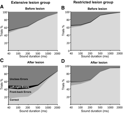
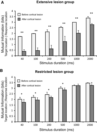
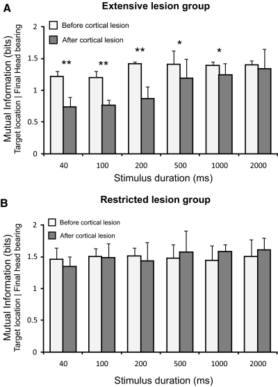
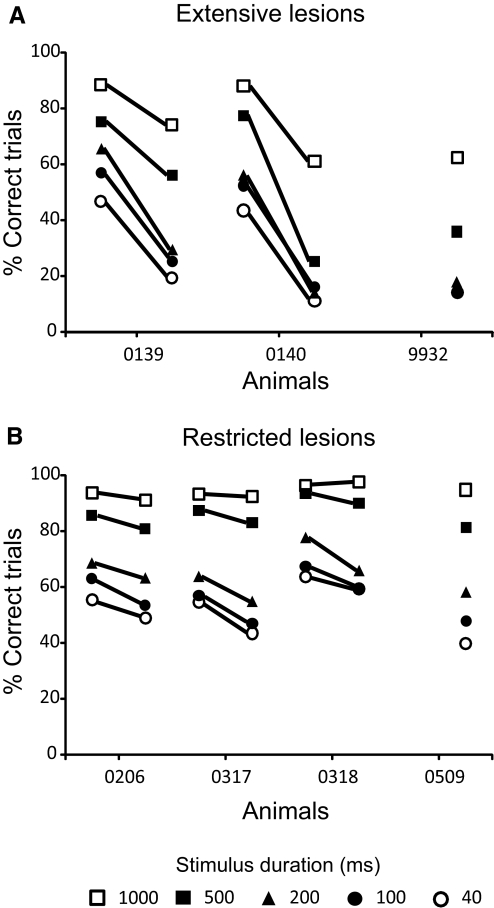
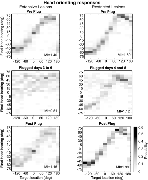
The role of auditory cortex in sound localization and its recalibration by experience was explored by measuring the accuracy with which ferrets turned toward and approached the source of broadband sounds in the horizontal plane. In one group, large bilateral lesions were made of the middle ectosylvian gyrus, where the primary auditory cortical fields are located, and part of the anterior and/or posterior ectosylvian gyrus, which contain higher-level fields. In the second group, the lesions were intended to be confined to primary auditory cortex (A1). The ability of the animals to localize noise bursts of different duration and level was measured before and after the lesions were made. A1 lesions produced a modest disruption of approach-to-target responses to short-duration stimuli (<500 ms) on both sides of space, whereas head orienting accuracy was unaffected. More extensive lesions produced much greater auditory localization deficits, again primarily for shorter sounds. In these ferrets, the accuracy of both the approach-to-target behavior and the orienting responses was impaired, and they could do little more than correctly lateralize the stimuli. Although both groups of ferrets were still able to localize long-duration sounds accurately, they were, in contrast to ferrets with an intact auditory cortex, unable to relearn to localize these stimuli after altering the spatial cues available by reversibly plugging one ear. These results indicate that both primary and nonprimary cortical areas are necessary for normal sound localization, although only higher auditory areas seem to contribute to accurate head orienting behavior. They also show that the auditory cortex, and A1 in particular, plays an essential role in training-induced plasticity in adult ferrets, and that this is the case for both head orienting responses and approach-to-target behavior.


Figures

Fig. 1.
Fig. 1.
Overall performance on the approach-to-target task for both groups before (A and B) and after (C and D) either extensive (A and C) or restricted (B and D) cortical lesions were made. Each panel shows the cumulative percentages of trials for the different sound durations used. Trials are sorted into those in which correct responses were made and, for the incorrect responses, are further subdivided into left-right errors (responses made to the hemifield contralateral to the target), front-back errors (responses made to the anterior hemifield following presentation of sounds in the ipsilateral posterior hemifield and vice versa), and unclassified (i.e., all other) errors. Note that the proportion of incorrect responses made by the animals in the 12-speaker task increased after the lesions had been made, particularly at shorter sound durations, but a much greater impairment was observed in the extensive lesion group.
Fig. 2.
Fig. 2.
Sound localization performance in the approach-to-target task for 2 representative cases (F0140, left column, which received extensive bilateral cortical lesions, and F0317, right column, which received smaller bilateral lesions that were restricted to the primary auditory cortex). Data were obtained at 2 different sound durations: 1,000 (A) and 40 ms (B). The size of the dots indicates, for each of the 12 target locations, the proportion of responses to different loudspeaker/reward-spout locations. For each plot, the value of the overall mutual information (MI) between response and target location is shown at the bottom right. A: localization performance at 1,000 ms was slightly impaired after extensive cortical lesions, but remained unaltered after restricted lesions. B: this difference was also apparent at 40 ms, where a modest disruption was seen after restricted cortical lesions and a much more substantial impairment was found after extensive cortical lesions.
Fig. 3.
Fig. 3.
The performance of the animals in the approach-to-target task was quantified by measuring the mutual information between the target and response locations. The mean and SD of these values are plotted across sound duration for the extensive lesion (A) and restricted lesion (B) groups both before (white bars) and after (gray bars) the cortex had been aspirated. In keeping with the change in the percentage of correct responses, the MI values were lower for shorter sound durations. The MI values were significantly reduced in both groups following the lesions, but a much larger effect was found in the extensive lesion group. **P < 0.01, *P < 0.05.
Fig. 4.
Fig. 4.
Effects of cortical lesions on sound-evoked head orienting responses. Final bearing of the initial head movement for the same representative cases shown in Fig. 2 (F0140 and F0317) in response to sounds of 2 different durations, 1,000 (A) and 40 ms (B), before and after the lesions were made. Each plot shows in grayscale the conditional probability of different final head bearings, with a bin size of 7.5°, for each target location. For each plot the value of the overall mutual information between final bearing and target location is shown at the bottom right. As with the approach-to-target responses, little effect was seen for long duration noise bursts (A), whereas for brief sounds, the clear correlation observed between the final head bearing and target location in the prelesion data was seriously disrupted after extensive cortical lesions, but only slightly affected after restricted lesions (B).
Fig. 5.
Fig. 5.
Mean and SD of the mutual information values between target location and final head bearing across sound duration for the extensive lesion (A) and restricted lesion (B) groups both before (white bars) and after (gray bars) the cortex had been aspirated. In contrast to the percent correct responses in the approach-to-target task, the MI values for the prelesion data were relative constant across sound duration. Extensive cortical lesions caused a significant reduction of the MI values for sounds of <500 ms in duration (A), whereas restricted cortical lesions did not alter the MI values (B). **P < 0.01, *P < 0.05.
Fig. 6.
Fig. 6.
Summary of the effects of extensive (A) or restricted (B) cortical lesions on sound localization accuracy. Mean prelesion and postlesion percent correct scores (averaged across all speaker locations) are shown for the individual animals in each group at different sound durations. Note the much larger drop in performance in the extensive lesion group. In 2 cases, equivalent prelesion data were not available because the animals had previously been used in a different auditory task. The postlesion scores of these animals were, however, very similar to those obtained for the other ferrets in each group.
Fig. 7.
Fig. 7.
Plasticity of sound localization depends on the auditory cortex. A: percent correct scores (averaged across all speaker locations) measured every day over a 10-day period. The performance is shown 1 day before (Pre) and on each of the 8 days after insertion of an earplug in the left ear (Left ear plugged), as well as on the day on which the earplug was removed (Post). The stimuli were 1,000-ms noise bursts. The gray bars depict the range of values obtained from 3 other ferrets with an intact auditory cortex. In these control animals, the percentage of correct responses falls dramatically when the ear is first plugged but recovers toward preplug levels with daily behavioral training. The different symbols correspond to the ferrets with cortical lesions, with the open symbols showing the data from the ferrets with restricted cortical lesions (the dashed line represents the mean scores for this group) and the filled symbols showing the data from 2 of the ferrets with extensive cortical lesions (the solid black line indicates the mean performance; the third animal in this group could not do the task with the earplug in place). In contrast to the controls, the localization abilities of the animals with cortical lesions did not improve during the period of time over which the earplug was worn. B: stimulus-response plots showing the approach-to-target performance before (Pre Plug) and after plugging the left ear (Plugged days 1–3 and Plugged days 7–9) and after removal of the earplug (Post Plug). The size of the dots indicates, for each of the 12 target locations, the proportion of responses to different loudspeaker/reward-spout locations, and the corresponding percent correct scores are given to the right of each panel. Monaural occlusion disrupted sound localization accuracy at all locations tested, but particularly on the side of the earplug. No improvement in performance was seen with daily localization training (middle panels). After earplug removal, sound localization accuracy returned to preplug levels.
Fig. 8.
Fig. 8.
Effect of monaural occlusion on acoustic head orienting behavior in the ferrets with cortical lesions. Each plot shows in grayscale the conditional probability of different final head bearings, with a bin size of 7.5°. These data were obtained from the same trials used to show the approach-to-target responses in Fig. 7. The correlation between final bearing and target location observed before inserting the ear plug (top row) was completely disrupted in the extensive lesion group when the left ear was occluded, with the animals turning toward the right side irrespective of the target location (left middle panel). A smaller bias to the right was also observed in the animals with restricted cortical lesions (right middle panel). In neither group did the accuracy of the orienting responses improve during the period of earplugging. Removal of the earplug resulted in a complete return to normal head orienting behavior (Post Plug).
Fig. 9.
Fig. 9.
Location and extent of the cortical lesions in the 3 animals (A, B, and C) classified as the “extensive lesion group.” For each case, a diagram of the cerebral cortex shows the lesioned area marked in gray, whereas areas in black represent where the underlying white matter was also aspirated. The coronal sections drawn at the level of the middle ectosylvian gyrus, as indicated by the line in A, show the total loss of the cortical layers and, in most cases, the underlying white matter. The approximate limits of the different auditory cortical areas described in the ferret are marked on a photomicrograph of the left ectosylvian gyri. AEG, anterior ectosylvian gyrus; ADF, anterior dorsal field; AVF, anterior ventral field; AAF, anterior auditory field; A1, primary auditory cortex; Hp, hippocampus; ls, lateral sulcus; LV, lateral ventricle; MEG, middle ectosylvian gyrus; PEG, posterior ectosylvial gyrus; PPF, posterior pseudosylvian field; PSF, posterior suprasylvian field; SSG, suprasylvian gyrus; sss, suprasylvian sulcus; pss, pseudosylvian sulcus; rf, rhinal fissure. Calibration bars represent 2 mm.
Fig. 10.
Fig. 10.
Degeneration in the thalamus after extensive lesions of the auditory cortex. Photomicrographs of coronal sections at the level of the medial geniculate body (MGB) and lateral geniculate nucleus (LGN) stained with a silver degeneration technique. Cortical lesions for this animal (F9932) are shown in Fig. 9A. A and B: low-power views of both sides of the brain. C–E: high-power photomicrograph of the different subdivisions of the right MGB. F: high-power photomicrograph of the right LGN. Squares in B represent the locations where each of these higher-magnification pictures was taken. Arrows in E and F indicate cells with a normal appearance that were observed in the medial division of the MGB and in the LGN. Calibration bars are 5 mm in A and B and 0.1 mm in F; calibration bar in F also applies to C–E.
Fig. 11.
Fig. 11.
Location and extent of the cortical lesions in the 4 animals in the “restricted cortical lesion” group. A: histological reconstructions of the cerebral cortex are shown schematically for each case. Note that the lesions were focused on the primary auditory cortex, which makes up only part of the MEG. In 2 animals, largely symmetrical lesions were made on each side; in the other 2, a greater proportion of the right cortex was aspirated, although the caudal part of the MEG, where A1 is located, was damaged on the left side also. In case F0509, the lesion also included part of the left suprasylvian gyrus. Coronal section in B shows for case F0317 that nearly all the cortical layers had been removed in the central part of the lesions and that there was virtually no encroachment on the underlying white matter. C: atrophy at the level of the A1 in the left hemisphere in cases F0318 and F0509. Compared with the control case shown in the left panel (F0640), the cortex is clearly damaged, with atrophy of the supragranular layers in F0318 (middle panel) and of all the layers in F0509 (right panel). Calibration bars are 2 mm in A and B and 0.5 mm in C. ps, pial surface; wm, white matter.

Similar articles

Cited by

References

    1. Bajo VM, Nodal FR, Bizley JK, Moore DR, King AJ. The ferret auditory cortex: descending projections to the inferior colliculus. Cereb Cortex 17: 475–491, 2007 - PMC - PubMed
    1. Bajo VM, Nodal FR, Moore DR, King AJ. The descending corticocollicular pathway mediates learning-induced auditory plasticity. Nat Neurosci 2009December27 [Epub ahead of print] - PMC - PubMed
    1. Beitel RE, Kaas JH. Effects of bilateral and unilateral ablation of auditory cortex in cats on the unconditioned head orienting response to acoustic stimuli. J Neurophysiol 70: 351–369, 1993 - PubMed
    1. Bizley JK, Nodal FR, Nelken I, King AJ. Functional organization of ferret auditory cortex. Cereb Cortex 15: 1637–1653, 2005 - PubMed
    1. Bizley JK, Nodal FR, Parsons CH, King AJ. Role of auditory cortex in sound localization in the midsagittal plane. J Neurophysiol 98: 1763–1774, 2007 - PMC - PubMed

Publication types

LinkOut - more resources