. 2010 Mar;298(3):R534-47.
doi: 10.1152/ajpregu.00367.2009. Epub 2009 Dec 23.

Overexpression of NGF in mouse urothelium leads to neuronal hyperinnervation, pelvic sensitivity, and changes in urinary bladder function

Affiliations

Overexpression of NGF in mouse urothelium leads to neuronal hyperinnervation, pelvic sensitivity, and changes in urinary bladder function

Birthe Schnegelsberg et al. Am J Physiol Regul Integr Comp Physiol. 2010 Mar.

Erratum in

  • Am J Physiol Regul Integr Comp Physiol. 2010 May;298(5):R1447

Abstract

NGF has been suggested to play a role in urinary bladder dysfunction by mediating inflammation, as well as morphological and functional changes, in sensory and sympathetic neurons innervating the urinary bladder. To further explore the role of NGF in bladder sensory function, we generated a transgenic mouse model of chronic NGF overexpression in the bladder using the urothelium-specific uroplakin II (UPII) promoter. NGF mRNA and protein were expressed at higher levels in the bladders of NGF-overexpressing (NGF-OE) transgenic mice compared with wild-type littermate controls from postnatal day 7 through 12-16 wk of age. Overexpression of NGF led to urinary bladder enlargement characterized by marked nerve fiber hyperplasia in the submucosa and detrusor smooth muscle and elevated numbers of tissue mast cells. There was a marked increase in the density of CGRP- and substance P-positive C-fiber sensory afferents, neurofilament 200-positive myelinated sensory afferents, and tyrosine hydroxylase-positive sympathetic nerve fibers in the suburothelial nerve plexus. CGRP-positive ganglia were also present in the urinary bladders of transgenic mice. Transgenic mice had reduced urinary bladder capacity and an increase in the number and amplitude of nonvoiding bladder contractions under baseline conditions in conscious open-voiding cystometry. These changes in urinary bladder function were further associated with an increased referred somatic pelvic hypersensitivity. Thus, chronic urothelial NGF overexpression in transgenic mice leads to neuronal proliferation, focal increases in urinary bladder mast cells, increased urinary bladder reflex activity, and pelvic hypersensitivity. NGF-overexpressing mice may, therefore, provide a useful transgenic model for exploring the role of NGF in urinary bladder dysfunction.


Figures
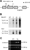
Fig. 1.
Fig. 1.
Generation of NGF-overexpressing (NGF-OE) transgenic mice. A: a 6,058 bp NotI fragment containing the uroplakin II (UPII)-NGFv2 transgene was microinjected into the pronuclei of fertilized C57BL/6J embryos to generate NGF-OE transgenic F0 lines. BamHI and Pst I restriction sites, and the position of the primers used for genotyping (RP41-NGF-S and βEx3-AS) are indicated. B: representative Southern blots of genomic DNA from WT and NGF-OE transgenic F0 lines 6, 20, and 23. Transgene integrations were characterized by BamHI digestion, which identified a 3.0-kb band from the endogenous (E) NGF gene and a predominant ∼6.1-kb band indicative of a head-to-tail arrangement of the stably integrated transgenes. Transgene copy number was estimated by Pst I digestion, which identified a 1.6-kb band from the single-copy endogenous (E) NGF locus and a 1.0-kb transgene band in the UPII-NGFv2 transgenic F0 lines. Densitometry scanning was used to estimate copy number as outlined in materials and methods. C: representative semiquantitative RT-PCR data showing amplification of total NGF mRNA isolated from the urothelium or detrusor smooth muscle bladder tissue of WT and NGF-OE transgenic mice. Total RNA was isolated and amplified as described in materials and methods and in Ref. . Amplicon size for total NGF mRNA is 268 bp.
Fig. 2.
Fig. 2.
NGF content in the urinary bladders of WT and NGF-OE transgenic mice. NGF content in whole urinary bladder was determined in WT and transgenic mice (F0 line 23) of different postnatal (P) ages, including P7-P10, 5–6 wk and 12–16 wk. Urinary bladder NGF was significantly (*P ≤ 0.01) greater in transgenic vs. WT mouse bladders at each postnatal age examined. Urinary bladder NGF content in transgenic mice was significantly (##P ≤ 0.01) greater in 12- to 16-wk-old mice compared with that detected in younger (P7-P10, 5–6 wk) transgenic mice. Urinary bladder NGF content in 5- to 6-wk-old transgenic mice was significantly (#P ≤ 0.01) greater than that detected in P7-P10 transgenic mice.
Fig. 3.
Fig. 3.
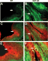
Colocalization of NGF and UPII in the urinary bladders of NGF-OE transgenic mice. Representative immunofluorescence labeling of NGF (green) (A, B) and representative coimmunofluorescence labeling of NGF (green) and UPII (red) in paraffin-embedded cross sections of urinary bladders (CF) from P7 WT (A, C, E) and transgenic (B, D, F) mice from F0 line 23. A: NGF immunoreactivity (green) was detected at low to moderate levels in the basal and intermediate cell layers of the bladder urothelium but was undetectable in the apical urothelial umbrella cells (arrow) of WT mice. B: in transgenic mice, NGF immunoreactivity was detected in all cell layers of the bladder urothelium, including a subpopulation of intermediate-to-apical urothelial cells (arrow), where it colocalized (yellow) with UPII (red) (D and F, arrows). UPII immunoreactivity (red) was most prominent in the apical cell layer of the bladder urothelium in both WT and transgenic mice, but it was also present in all suprabasal cell layers of the urothelium (CF). E and F are a higher magnification of the images shown in C and D. An NGF-blocking peptide completely abolished the staining for NGF (not shown). Scale bar: 50 μm (AD), 25 μm (E, F).
Fig. 4.
Fig. 4.
Histopathology of the urinary bladders of WT and NGF-OE transgenic mice. Representative histology images of Masson Trichrome-stained cross sections of the urinary bladders of 8-wk-old female WT (A, C) and transgenic (B, D) mice from F0 line 23 (n = 3–5). The lumen (L), urothelium (U), and detrusor smooth muscle (DSM) layers are indicated. The Masson Trichrome stain is well suited for distinguishing epithelial vs. surrounding connective tissue structures, staining keratin and muscle fibers red, collagen fibers blue, and nerve fibers pale pink. Note the DSM and U in red and the suburothelium in blue in WT and transgenic urinary bladders (A, B) and the marked expansion of nerve fiber tissue in the suburothelium of transgenic mice (B, see arrows; D), appearing here as a pale pink stain. Scale bar: 100 μm (A, B), 50 μm (C, D).
Fig. 5.
Fig. 5.
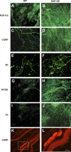
Neuronal hyperinnervation in the urinary bladders of NGF-OE transgenic mice. Representative fluorescence images of PGP 9.5 (A, B), CGRP (C, D), Substance P (E, F), NF200 (G, H), and TH (I, J) immunoreactivity in the bladder neck region in urothelial whole-mount preparations of urinary bladders from 8- to 10-wk-old female WT (A, C, E, G, I) and NGF-OE transgenic (B, D, F, H, J) mice from F0 line 23 (n = 3–5). (K, L) Representative fluorescence images of CGRP-immunoreactive ganglia interspersed with CGRP-immunoreactive nerve fibers in urothelial whole mount preparations from NGF-OE transgenic mice. Dashed box in K is expanded in higher power in L. Scale bar: 50 μm (AJ), 80 μm (K), and 40 μm (L). Protein Gene Product (PGP 9.5), calcitonin gene-related protein (CGRP), substance P (SP), neurofilament (NF 200), tyrosine hydroxylase (TH).
Fig. 6.
Fig. 6.
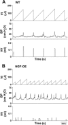
Open cystometry in conscious WT and NGF-OE transgenic mice. Representative cystometrogram trace from conscious, unrestrained WT (A) and NGF-OE (B) mice from F0 line 23 during a continuous intravesical infusion (25 μl/min) of room temperature saline. Volume infused (VI, μl), bladder pressure (BP, cm H2O) and voided volume (VV, ml) are shown. Arrows indicate examples of nonvoiding bladder contractions.
Fig. 7.
Fig. 7.
Summary bar graphs from open cystometry. Bar graphs of VV (A, ml), ICI (B, s) and BP (C, cm H2O) from open cystometry in conscious, unrestrained WT and NGF-OE transgenic mice from F0, line 23. VV (A) and ICI (B) were significantly (*P ≤ 0.001) reduced in transgenic mice compared with WT mice. C: no changes in baseline, threshold, or maximum micturition pressure were observed between WT and transgenic mice during conscious cystometry. Data represent the mean ± SE of n = 10–12 mice per group.
Fig. 8.
Fig. 8.
Nonvoiding urinary bladder contractions (NVCs) in NGF-OE transgenic mice from open cystometry. Representative cystometrogram trace from a conscious, unrestrained transgenic mouse from F0 line 23 during continuous intravesical infusion (25 μl/min) of room temperature saline. VI (A, μl) and BP (B, cm H2O) are shown. Dashed box in B draws attention to two micturition events associated with the occurrence of NVCs. C: dashed box in B is expanded with arrows indicating NVCs and asterisks indicating void events.
Fig. 9.
Fig. 9.
Summary bar graphs of NVC properties in WT and NGF-OE transgenic mice. NVCs were observed in both WT and transgenic mice under conscious, open cystometry conditions but were not observed with each voiding cycle in either WT or transgenic mice. The number of NVCs/voiding cycle (A), amplitude of NVCs (B, cm H2O), and frequency of occurrence of NVCs/voiding cycle (C, %) were significantly (*P ≤ 0.01) increased in transgenic mice. Data represent the means ± SE of n = 10–12 mice per group.
Fig. 10.
Fig. 10.
Somatic sensitivity testing in WT and NGF-OE transgenic mice. Pelvic region testing with calibrated von Frey hairs was determined in WT and NGF-OE transgenic mice from F0 line 23. The von Frey hairs were applied in an up-down method for 1–3 s with an interstimulus interval of 15 s. For pelvic region stimulation, stimulation was confined to the lower abdominal area overlying the urinary bladder. The following behaviors were considered positive responses to pelvic region stimulation: sharp retraction of the abdomen, jumping, or immediate licking or scratching of the pelvic area (90). Transgenic mice had a significantly (*P ≤ 0.001) increased pelvic response frequency with all von Frey hairs (0.1–4 g) tested compared with WT mice. Hindpaw sensitivity testing with calibrated von Frey hairs was determined in separate groups of WT and NGF-OE transgenic mice. No differences in the paw pressure threshold required to elicit a sharp withdrawal of the paw or licking of the tested hindpaw (90) were detected between WT and transgenic mice. Data represent the mean ± SE of n = 8 mice per group. All somatic testing and behavioral scoring were performed in a blinded manner with respect to treatment and mouse strain. Pts., points.

Comment in

Similar articles

Cited by

References

    1. Alagiri M, Chottiner S, Ratner V, Slade D, Hanno PM. Interstitial cystitis: unexplained associations with other chronic disease and pain syndromes. Urology 49: 52–57, 1997 - PubMed
    1. Albers KM, Wright DE, Davis BM. Overexpression of nerve growth factor in epidermis of transgenic mice causes hypertrophy of the peripheral nervous system. J Neurosci 14: 1422–1432, 1994 - PMC - PubMed
    1. Allen SJ, Dawbarn D. Clinical relevance of the neurotrophins and their receptors. Clin Sci 110: 175–191, 2006 - PubMed
    1. Aloe L. The effect of nerve growth factor and its antibody on mast cells in vivo. J Neuroimmunol 18: 1–12, 1988 - PubMed
    1. Andreev NY, Dimitrieva N, Koltzenburg M, McMahon SB. Peripheral administration of nerve growth factor in the adult rat produces a thermal hyperalgesia that requires the presence of sympathetic post-ganglionic neurones. Pain 63: 109–115, 1995 - PubMed

Publication types

MeSH terms